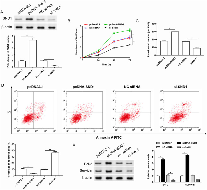
Figure 4.
Overexpression of SND1 promoted proliferation and invasion, and inhibited apoptosis in cervical cancer cells. SND1 was overexpressed or silenced in HeLa cells. (A) The transfection efficiency was detected by western blotting, and the quantification was performed using Image J software. n = 3. *P < 0.05, **P < 0.01, one-way ANOVA. (B-D) The cell proliferation, invasion, and apoptosis were evaluated by Cell Counting Kit 8, Transwell, and Annexin V-FITC/PI assay, respectively. n = 3. *P < 0.05, **P < 0.01, one-way ANOVA. (E) The expression of anti-apoptotic proteins was detected by western blotting, and the quantification was performed using Image J software. n = 3. *P < 0.05, one-way ANOVA.
ANOVA: analysis of variance; FITC: fluorescein isothiocyanate; NC: negative control; PI: propidium iodide.

