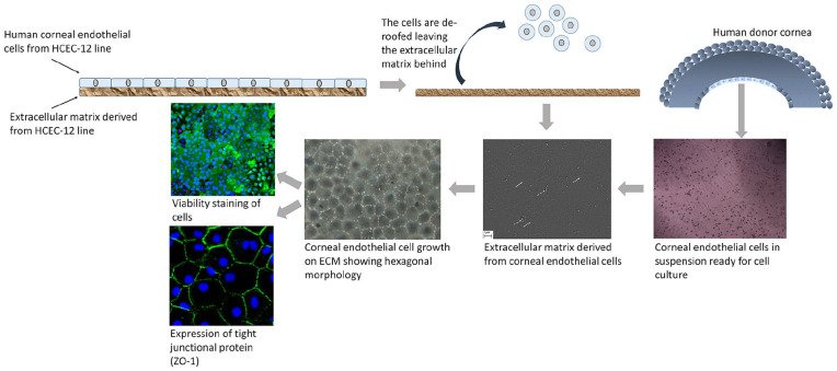
Figure 3.
Human corneal endothelial cell culture on HCEC-12 derived extracellular matrix (ECM). HCEC-12 cells are cultured on a culture plate. ECM is laid by the HCEC-12 cells naturally. Upon confluence, the cells are detached leaving behind the ECM. Corneal endothelial cells from human donor corneas are isolated and cultured on the ECM. White arrows show fiber-like collagen structures and dotted white arrow show cell debris. Cell morphology, viability and expression of tight-junction protein are checked to confirm the health and for end-stage characterization.

