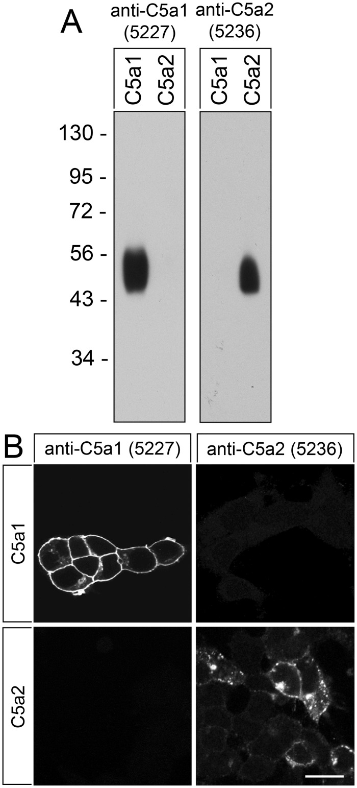
Fig 2. Analysis of the specificity of anti-human rabbit polyclonal antibody {5227}.
(A) Western blot analysis of whole cell preparations from stably C5aR1- or C5aR2-transfected HEK-293 cells. Receptors were enriched using wheat germ lectin agarose beads. Samples were separated on 7.5% SDS-polyacrylamide gels and blotted onto PVDF membranes. Membranes were then incubated with anti-human C5aR1 antibody {5227} or anti-human C5aR2 antibody {5236}, and blots were developed using enhanced chemiluminescence. Ordinate, migration of protein molecular weight markers (kDa). Note that {5227} and {5236} selectively detected only the respective targeted receptor and did not cross-react with the other receptor or other membrane proteins. (B) Immunocytochemistry of stably C5aR1- or C5aR2-transfected HEK-293 cells. Cells were fixed and immunofluorescently stained with anti-human C5aR1 antibody {5227} or anti-human C5aR2 antibody {5236}. Again, {5227} and {5236} selectively detected only the targeted receptor and did not cross-react with the other receptor. Note also, that prominent immunofluorescence for C5aR1 was localised at the plasma membrane. Representative results from one of three independent experiments are shown. Scale bar: 20 μm.

