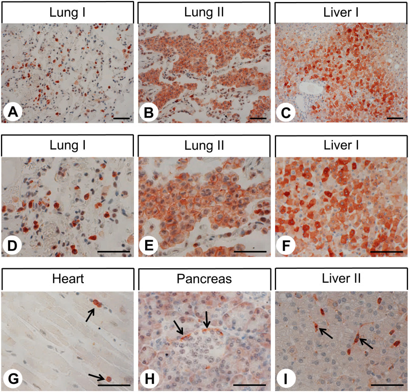
Fig 3. Immunohistochemical detection of C5aR1 localisation in non-neoplastic human tissues (I).
Immunohistochemical staining (red-brown colour), counterstaining with haematoxylin. Scale bar: 100 μm (C, F), 50 μm (A, B, D, E, G-I). Arrows in (G): positive immune cells in the heart; arrows in (H): positive cells at the margin of the pancreatic islet; arrows in (I): positive Kupffer cells in the liver. (D-F) enlarged sections of (A-C).

