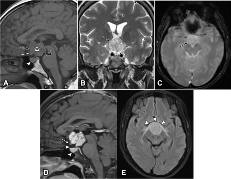
Figure 2.
Brain MRI of a 46-year-old woman, showing a lobulated, contoured, solid mass. (A) Mid-sagittal T1-weighted image showing a hypointense lesion (white star), expanding into the supraoptic recess (black arrow), bowing the floor of the third ventricle (curved arrow), pushing the optic chiasm downward and forward (white arrow), and the pituitary gland remains intact (white arrowhead). (B) Coronal T2-weighted image showing a heterogeneous, hyperintense lesion (arrow). (C) Axial T2-weighted image showing no intratumoral calcification (arrow). (D) Sagittal T1-weighted image, post-contrast, showing vivid heterogeneous enhancement. The optic chiasm is compressed downward and forward (white arrow), and the suprasellar cistern and the pituitary gland are intact (arrowheads). (E) Axial fluid-attenuated inversion recovery (FLAIR) image showing the anterior deviation and edema of the optic chiasm (arrowheads).

