
Figure 7-30.
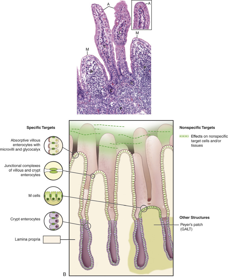
Targets for Microbial Infection in the Intestine.
A, Photomicrograph of small intestinal mucosae identifying targets for infection. Compare with schematic diagram illustrated in B.Inset, Higher magnification of villus tip enterocytes with a microvillus border. B, Schematic diagram illustrating targets for infection. A, Absorptive enterocyte; C, undifferentiated crypt cells; GALT, gut-associated lymphoid tissue; L, lamina propria: M, M cells; P, Peyer's patch.
(A and inset courtesy Dr. J.F. Zachary, College of Veterinary Medicine, University of Illinois.)