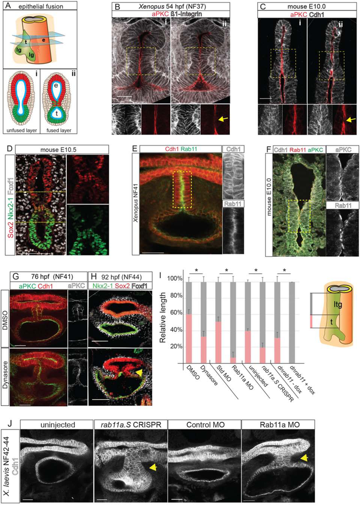
Figure 2: Endosome-Mediated Epithelial Remodeling is Required for TE Septation.
2A: Model of epithelial fusion.
2B–C: Sequential optical sections of X. laevis (B) and mouse (C) embryo immunostaining showing loss of aPKC and increased Integrin or Cdh1 at the contact point (arrow, ii). Scale bar, 50 μm.
2D: A unique population of cells co-expressing of Sox2 and Nkx2–1 in the mouse foregut. Scale bar, 100 μm.
2E: Rab11 and Cdh1 are enriched in the X. laevis septum. Scale bar, 50 μm.
2F: Rab11, aPKC and Cdh1 enriched at the fusion point in mouse. Scale bar, 50 μm.
2G–H: Inhibition of endosome recycling by Dynasore treatment of X. laevis (NF32–41) results in a failure to reduce aPKC at NF41 (G) and a TEC at NF44 (H). Scale bar, 100 μm.
2I: Quantification of reduced trachea (t) length and relative to laryngotracheal (ltg) in NF42–44 X. laevis embryos. Student’s two-tailed t-test, between manipulated and control sibling embryos *p<0.05.
2J. Rab11a CRISPR-mediated mutation or MO knockdown results in a TEC at NF42–44 in X. laevis. Scale bar, 50 μm.

