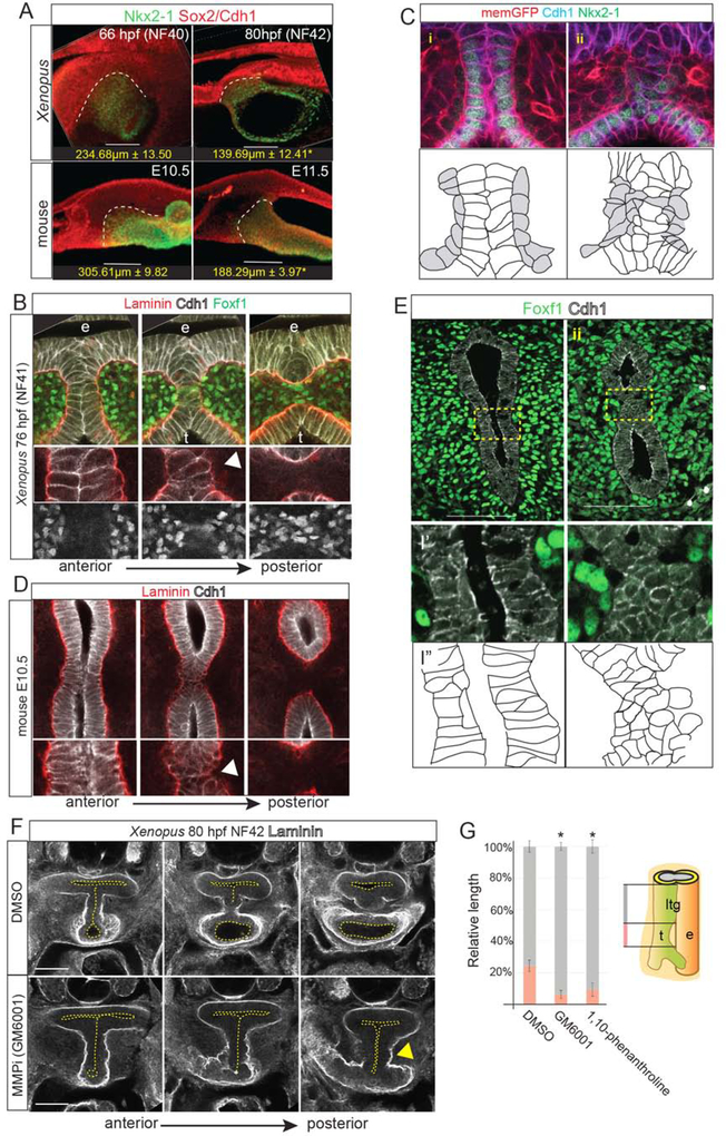
Figure 3: Localized ECM Degradation and Mesenchymal Invasion Resolve the TE Septum.
3A: Wholemount immunostaining of septum resolution in X. laevis and mouse embryos, quantifying the length of the Sox2+/Nkx2–1+ boundary. Student’s two-tailed t-test,*p<0.05.
3B: Serial optical sections showing Laminin, Cdh1 and Foxf1 during TE septum resolution in NF41 X. laevis embryos. Arrowhead indicates localized Laminin breakdown.
3C: Immunostaining of transgenic membrane GFP NF41 Xenopus embryo (i) anterior to and (ii) at the septation point showing Cdh1+ epithelial (white in schematic) round up as mesenchymal cells (grey) invade. Scale bar, 100 μm.
3D: Serial optical sections showing Laminin and Cdh1 during TE septum resolution in E10.5 mouse embryos. Arrowhead indicates localized Laminin breakdown.
3E: Immunostaining of Foxf1 and Cdh1 (schematic below) in an E10.5 mouse embryo (i) anterior to and (ii) at the septation point. Scale bar, 100 μm.
3F: Inhibition of MMP activity in Xenopus with GM6001 (from NF32–42) results in impaired Laminin breakdown (arrowhead) and a TEC.
3G: Quantification of relative lengths of the laryngotracheal groove (ltg) and trachea (t) in DMSO, GM6001, or 1,10-phenanthroline-treated NF41 X. laevis embryos. One-way ANOVA, *p<0.05.

