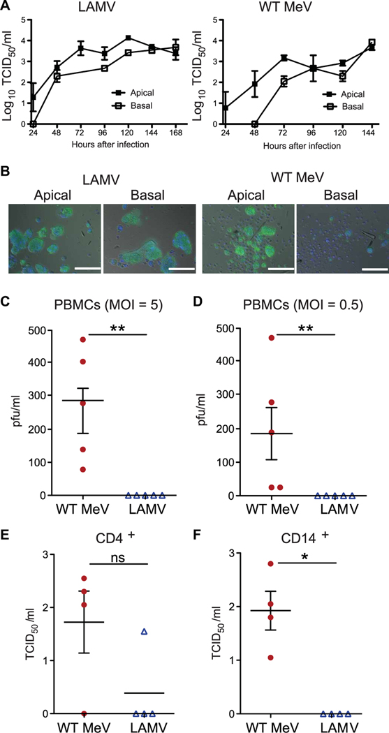
Fig. 4. In vitro replication of WT MeV and LAMV in primary respiratory epithelial cells and PBMCs.
Infectious MeV in apical supernatants and shedding of MeV-infected multinuclear giant cells were measured after apical or basal infection of primary differentiated macaque tracheal epithelial cells with LAMV or WT MeV (MOI = 4.5). (A) Apical cell culture supernatants (cultures from three separate macaques; two replicates in each experiment) were assayed for infectious virus by a TCID50 assay. Lines indicate the SEM. (B) Shed cells collected from macaque tracheal epithelial cell monolayers 144 hours after infection were stained with antibody to MeV N protein and DAPI nuclear stain after fixation and permeabilization. Merged images of phase-contrast, MeV N protein expression (green), and DAPI nuclear stain (blue) are shown. Scale bar represents 150 μm. (C to F) Production of infectious virus 24 hours after infection of human PBMCs with WT MeV (red circles) and LAMV (blue triangles) at high (2 to 5) MOI (C) and low (0.5) MOI (D). Production of infectious virus 24 hours after infection of human CD4+ T cells (E) or CD14+ myeloid cells (F) with WT MeV (red circles) or LAMV (blue triangles) (MOI = 5). Horizontal line indicates the mean. Significance was determined by Mann-Whitney U test. *P < 0.05 and **P < 0.01. ns, not significant.

