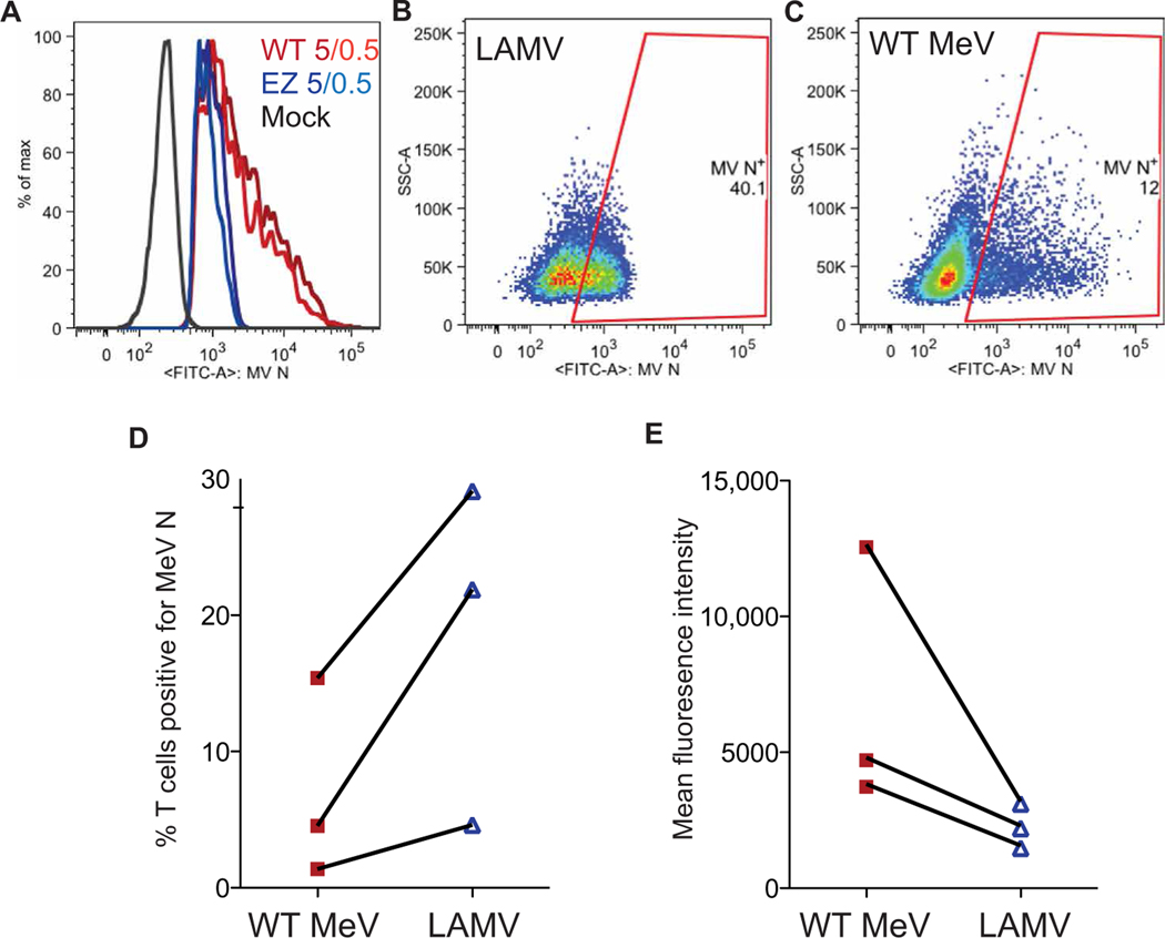
Fig. 5. In vitro replication of WT MeV and LAMV in human primary CD4+ T cells.
Shown is flow cytometry analysis of MeV N protein expression after in vitro infection of human PBMCs with WT MeV or LAMV (vaccine strain of MeV). (A) Flow cytometry histogram shows amounts of MeV N protein expressed by human CD4+ T cells 20 hours after infection with WT MeV or LAMV at an MOI of 0.5 or 5.0. (B and C) Flow cytometry plots of CD4+ T cell expression of MeV N protein 20 hours after infection with LAMV (B) or WT MeV (C) strains (MOI = 5.0). SSC, side-scattered light. (D and E) Each panel shows results of three separate flow cytometry experiments that assessed the percentage of human CD4+ T cells expressing MeV N protein (D) and the amount of MeV N protein measured by immunofluorescence (E) 48 hours after infection of human PBMCs with WT MeV or LAMV (MOI = 5). Lines connect data from the same experiment.

