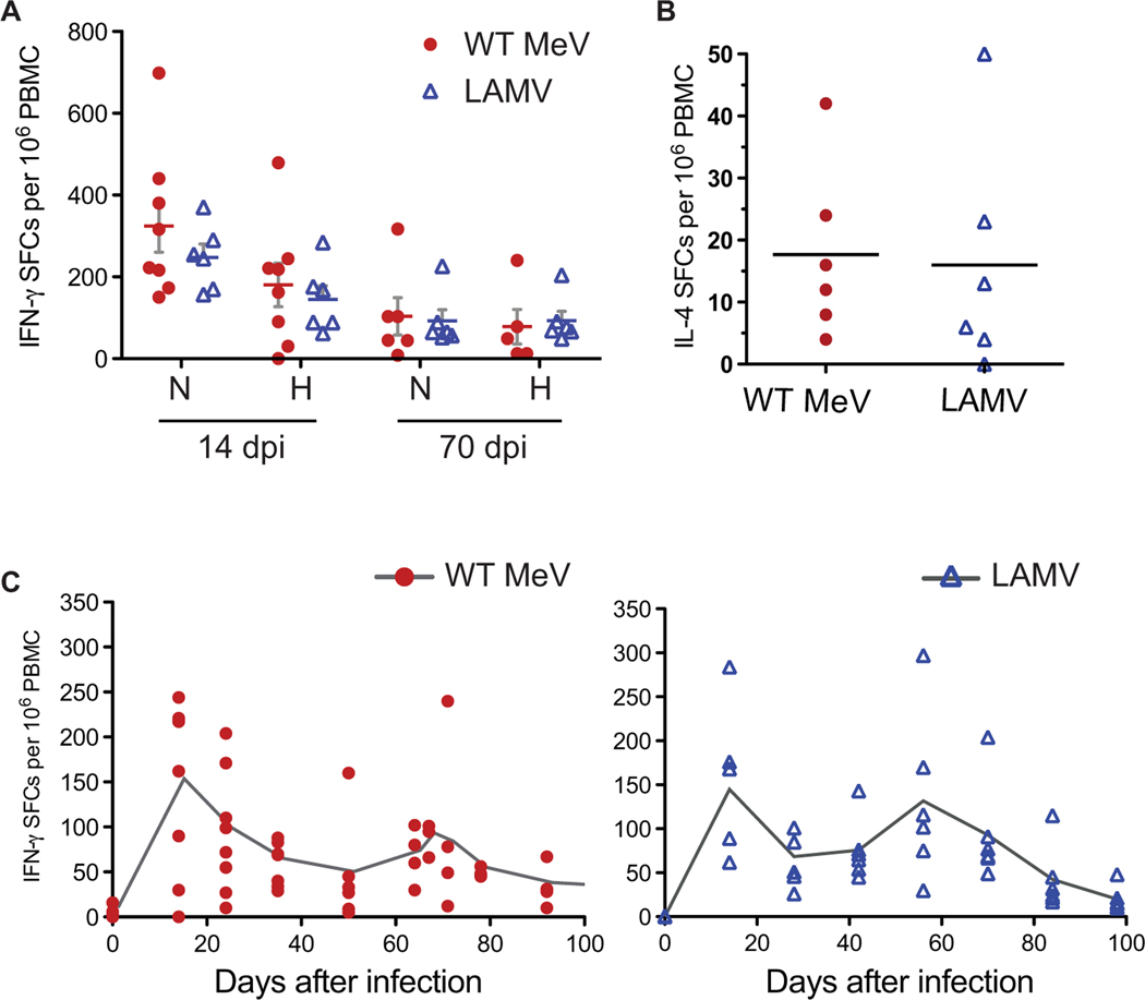
Fig. 8. Cellular immune responses after WT MeV and LAMV infection of macaques.
Numbers of IFN-γ– and IL-4–producing T cells in infected macaques were assessed by ELISpot assay. Fresh macaque PBMCs (5 × 10⁵) were added to multiscreen plates coated with anti-human IFN-γ or IL-4 antibody in the presence of pooled H or N peptides or medium. Data are reported as spot-forming cells (SFCs) after subtracting medium alone wells from antigen-stimulated wells. (A) Numbers of H- and N-specific IFN-γ–producing cells 14 and 70 days postinfection (dpi). (B) Numbers of N-specific IL-4–producing cells 14 days after infection. Horizontal line indicates the mean. (C) Changes in numbers of N-specific IFN-γ–producing cells in the circulation of infected macaques over the course of MeV infection. Reactivation of MeV-specific T cells in the circulation was noted around 60 days after infection for animals infected with WT MeV (red circles) or LAMV (blue triangles).

