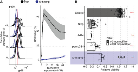
Fig. 5. Contribution of p38 to apoptosis in hypertonic stress is minimal.

(A) Phosphorylation of p38 in Jurkat cells exposed to 300 mosmol/liter NaCl by a step (black) or a 10-hour ramp (blue). The left panel shows selected single-cell distributions over the cumulative exposure with individual lines representing independent experiments. The red line indicates the threshold for determining a cell that is p38 phosphorylation positive (ON-fraction). The right panel represents the ON-fraction mean and SD of 3 to 10 independent experiments as a function of cumulative exposure. (B) Viability of Jurkat cells relative to untreated cells (control) exposed to an additional 0 (gray) or 300 mosmol/liter (white) NaCl for 5 hours (step) or 10 hours (ramp, purple), respectively. Pan p38 inhibitor (pan-p38-i, BIRB796, 10 μM) and JNK inhibitor (SP600125, 10 μM) were added 30 min before NaCl. Circles represent single experiments. Bars indicate the mean and SD of at least three replicates. Two-sided unpaired Student’s t test: ***P < 0.01 and ****P < 0.0001.
