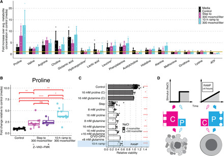
Fig. 6. Intracellular proline protects human cells during ramp stress conditions.

(A) Fifteen most abundant metabolites detected in Jurkat cells without stimulation (black), after treatment with step (magenta) or 10-hour ramp (cyan) to 300 mosmol/liter NaCl. Bars represent mean and SD of the fold change of each metabolite to the average metabolite concentration in the control condition (yellow line) with circles representing replicates. (B) Change of proline levels in Jurkat cells relative to control (no additional NaCl) in 0 (black) or step without (magenta) or with pan-caspase inhibitor “a” (Z-VAD-FMK, 100 μM) (purple) or 10-hour ramp to 300 mosmol/liter NaCl (cyan). Circles represent replicates, N ≥ 4. (C) Viability in Jurkat cells exposed to step or 10-hour ramp (blue) of 300 mosmol/liter NaCl relative to control (gray). Pan-caspase inhibitor (Q-VD-OPH, 100 μM) was added 30 min and amino acids 60 min before NaCl. Bars indicate mean and SD, N ≥ 3. Two-sided unpaired Student’s t test: *P < 0.05, **P < 0.01, ***P < 0.001, and ****P < 0.0001. (D) Model: Instant stress conditions cause activation of caspase signaling (C) and cell death (magenta), whereas the gradual increased stress to the same final concentration does not activate caspase signaling but instead increases intracellular proline (P) as an osmolyte to protect cells against increasing stress (cyan).
