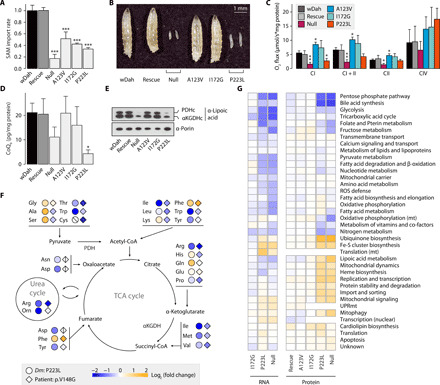
Fig. 1. Acute mitoSAM depletion is lethal through loss of mitoSAM-dependent metabolites.

(A) SAM import rates into larval mitochondria (n = 3) relative to the genetic background control wDah. (B) Four-day-old mutant Dm larvae. (C) Mitochondrial oxygen consumption rates in larvae (n = 5) normalized to protein content. (D) CoQ9 levels in larval mitochondria (n = 3) normalized to protein content of input larvae. (E) Immunoblot on mitochondrial lysates from larvae, against lipoylation of pyruvate dehydrogenase (PDHc) and α-ketoglutarate dehydrogenase (αKGDHc) subunit E2, or porin as the loading control. (F) Total amino acid levels in p223l larvae relative to wDah controls (n = 3) or in serum of the previously reported p.V148G patient relative to mean standard values [diamonds; patient 1 in (9)]. Crossed shapes, metabolite not detected. CoA, coenzyme A. TCA, tricarboxylic acid. (G) Mitochondrial subsetting of larval RNA sequencing and proteomics data (n = 5). Expression level relative to wDah controls and vertical hierarchical clustering. UPRmt, mitochondrial unfolded protein response. Bar graphs show means + SD. *P < 0.05, **P < 0.01, and ***P < 0.001 with Dunnett’s test against wDah (A, C, and D). All experiments were performed on 4-day-old Dm larvae.
