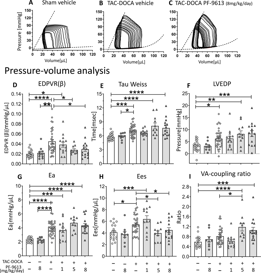
Figure 2. Pressure-Volume analysis of TAC-DOCA mice.
Representative PV analysis of sham-veh (A), TAC-DOCA-veh (B) and TAC-DOCA-inh at PF-4449613 concentration of 8 mg/kg/day (C). TAC-DOCA-veh mice exhibit increased LV chamber stiffness (EDPVR (β)) (D), prolonged relaxation (Tau) (E), elevated LV diastolic filling pressure (LVEDP) (F), increased effective arterial elastance (Ea)(G) and increased LV contractility (Ees) (H). The EDPVR (β) of TAC-DOCA-inh1 is not different from TAC-DOCA-veh, but the EDPVR (β) of TAC-DOCA-inh5 and TAC-DOCA-inh8 are lower compared to TAC-DOCA-veh (D). However LVEDP of TAC-DOCA-inh5 and TAC-DOCA-inh8 remains elevated (F) and the VA coupling ratio increases, suggesting a decline in VA co-ordination (I). EDPVR indicates End diastolic pressure volume relation; Tau, relaxation time constant; Ees, End systolic elastance; LVEDP, left ventricular end diastolic pressure; VA co-ordination, ventricular-arterial coordination. (n= 22, 10, 33, 14, 11, 15 mice) * p≤0.05 ** p≤0.01 ***p≤0.001 ****p≤0.0001. Two-way ANOVA, without repeated measures, with a Dunnett test was used.

