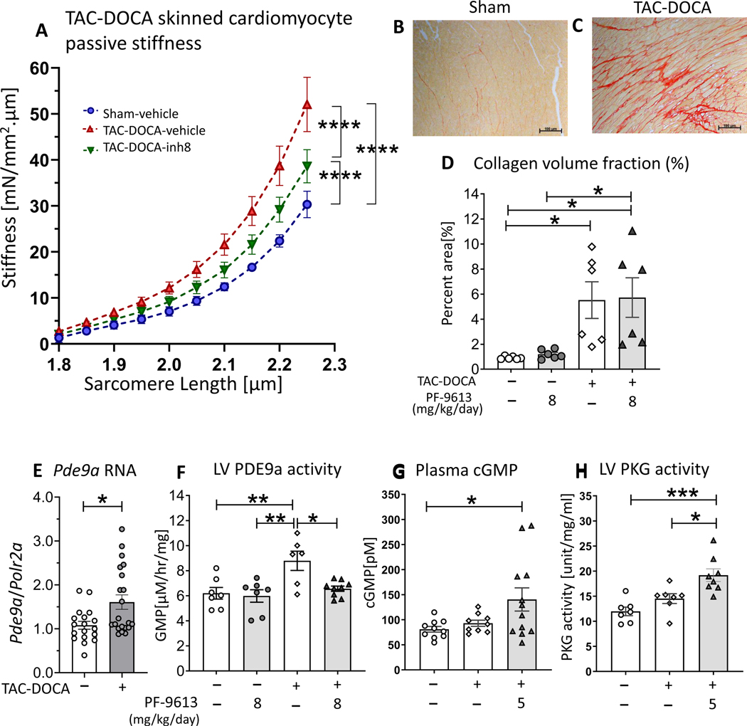
Figures 4. Cardiomyocyte passive stiffness, LV collagen content, LV Pdea9 RNA expression, LV PDE9a activity, plasma cGMP and LV PKG activity of TAC-DOCA mice.
Cardiomyocyte passive stiffness after chronic PDE9a inhibition, measured in demembraned (skinned) LV cardiomyocytes (A). Cellular stiffness is increased in both groups of TAC-DOCA mice, however, the stiffness of TAC-DOCA-inh8 is lower than TAC-DOCA-veh. (n= 7,15,14 cells from 4,8,7 mice) (A). Representative Picrosirius Red staining for collagen of LV myocardium (B&C). Quantitative analysis shows a significant increase in percent area of myocardial collagen in both groups of TAC-DOCA mice (D). No difference in percent area of collagen is observed between TAC-DOCA-veh and TAC-DOCA-inh8 (n=7, 6, 6 and 7 mice). Quantitative PCR was used to assess Pde9a RNA expression in LV myocardium. There is a significant upregulation of Pde9a mRNA at 5 weeks after TAC-DOCA surgery (E) (n=18,21 mice). High performance liquid chromatography was used to assess PDE9a activity in LV myocardium. PDE9a activity is increased in TAC-DOCA-veh but is normalized in TAC-DOCA-inh8 (n=7,7,6,9 mice) (F). Plasma concentration of cGMP (G) and LV myocardial PKG activity (H) are increased in TAC-DOCA mice with PDE9a inhibition (n=10,9,12 mice for plasma cGMP and n=7,7,8 mice for PKG activity). * p≤0.05 ** p≤0.01 ***p≤0.001 ****p≤0.0001. Statistical analyses consisted of: (A) Nonlinear regression analysis with a least squares fitting, (D&F) Two-way ANOVA, without repeated measures, with a Tukey test, (E) Mann-Whitney U test, (G) Kruskal-Wallis with a Dunn test,(H) One-way ANOVA without repeated measures with a Tukey test.

