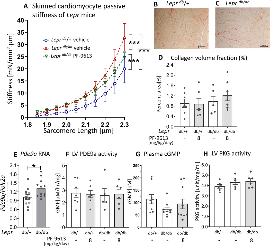
Figure 8. Cardiomyocyte passive stiffness, LV collagen content, LV Pde9a RNA expression, LV PDE9a activity, plasma cGMP and LV PKG activity of Leprdb/db mice.
Cardiomyocyte passive stiffness after chronic PDE9a inhibition, measured in demembraned (skinned) LV cardiomyocytes (A). Cardiomyocyte stiffness is increased in both groups of Leprdb/db mice, however, the stiffness is slightly reduced in the Leprdb/db mice that were treated with PF-4449613 compared to vehicle (A) (n= 3,16,15 cells from 2,6,7 mice). Representative Picrosirius Red staining for collagen of LV myocardium (B&C). Quantitative analysis does not show a significant difference in percent area of collagen among groups (n=7, 6, 6 and 7 mice) (D). There is a significant upregulation of Pde9a mRNA (n=12,12 mice) (E), however, there is no increase in PDE9a activity in LV myocardium of Leprdb/db mice (F) (n= 7,7,5,6 mice). There is no increase in plasma cGMP concentration (G) or LV PKG activity (H) in Leprdb/db mice with PDE9a inhibition (n =9,9,11 mice for plasma cGMP and n = 6,6,6 mice for LV PKG activity), * p≤0.05** p≤0.01 ***p≤0.001 ****p≤0.0001. Statistical analyses consisted of: (A) Nonlinear regression analysis with a least squares fitting, (D&F) Two-way ANOVA, without repeated measures, with a Tukey test, (E) unpaired t test, (G) Kruskal-Wallis with Dunn test, (H) One-way ANOVA without repeated measures with a Tukey test.

