. 2021 Feb 8;10:e62326. doi: 10.7554/eLife.62326

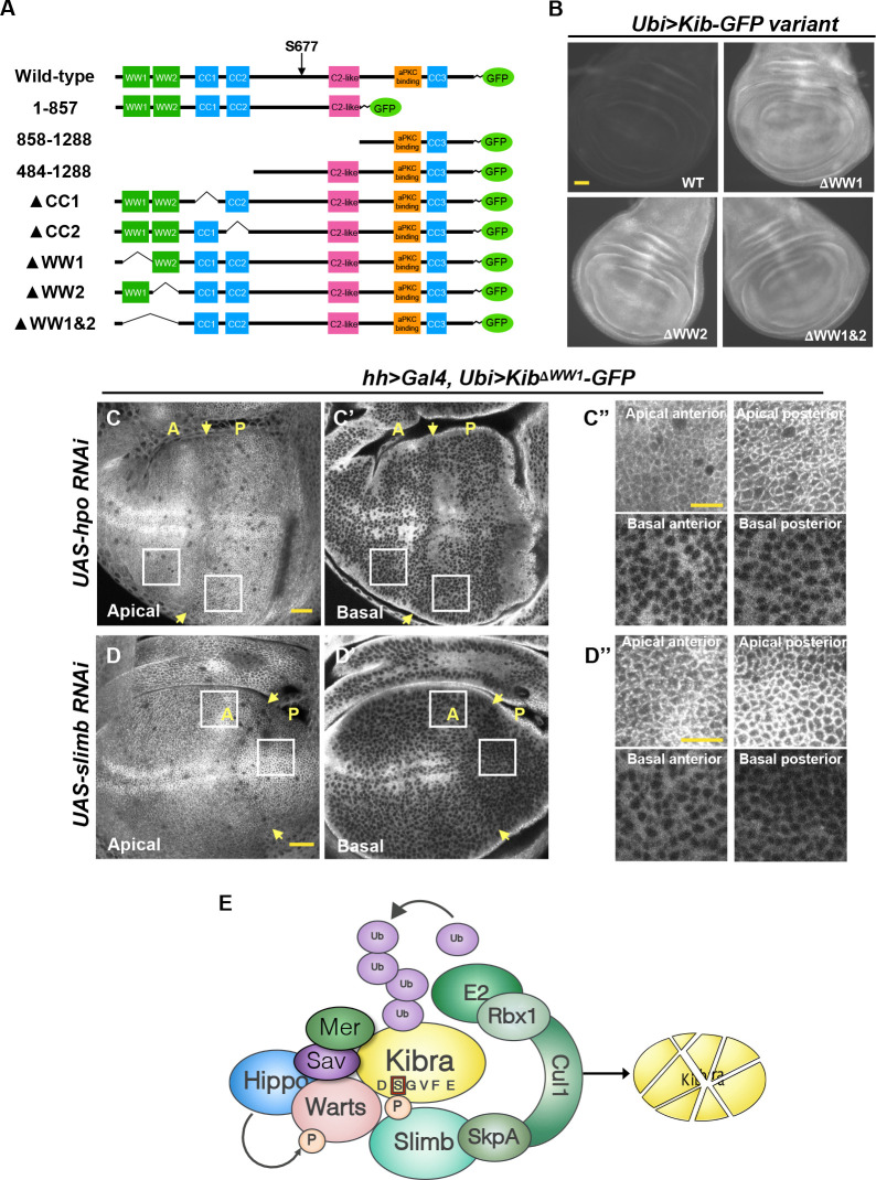
Figure 6. The WW domains of Kibra (Kib) are required for Hippo (Hpo) pathway- and Slimb-mediated degradation.

(A) Diagram of Kib truncations generated for this study. (B) Widefield fluorescence images of wing imaginal discs expressing wild-type and WW-domain truncations of Kib-GFP expressed under the ubiquitin promoter. All images were taken with identical settings. Scale bar=40 μm. (C–C’’) Depletion of Hpo does not affect expression of Ubi>KibΔWW1-GFP. Note that Hpo depletion leads to apical stabilization and basal depletion of KibΔWW1-GFP (C’’). (D–D’’) Depletion of Slimb does not affect expression of Ubi>KibΔWW1-GFP. Note that similar to Hpo depletion, loss of Slimb leads to slight apical stabilization and basal depletion of KibΔWW1-GFP (D’’). Yellow arrows indicate A–P boundary of the wing discs. Scale bars=20 μm (C and D) and 10 μm (C’’ and D’’). (E) A model of Kib degradation by the Hpo pathway and Slimb.

Figure 6.

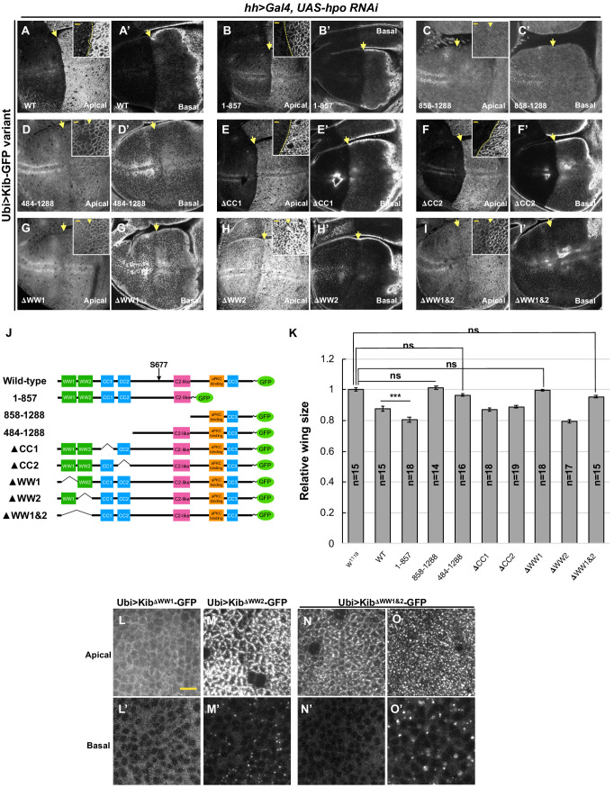
Figure 6—figure supplement 1. The role of WW domains in Hippo pathway-mediated Kibra (Kib) degradation.

Figure 6—figure supplement 1.

(A–I’) Effect of Hpo depletion in the posterior compartment of the wing imaginal disc on different Kib truncations. Deletion of the WW domains, individually (G–H’) or together (I and I’) stabilizes Kib apically but does not lead to an increase in basal Kib levels. Note that tissue in (G) is the same as shown in Figure 6C. Scale bars in the insets represent 3 μm for the insets and 10 μm for the corresponding low magnification image. (J) Diagram of Kib truncations generated for this study (same as in Figure 6A). (K) Size comparison (relative to wild-type) of adult wings from flies ectopically expressing different Ubi>Kib-GFP truncations. Quantification is shown as the mean ± SEM; n=number of wings. Statistical comparison was performed using the one-way ANOVA test followed by Tukey’s HSD test. (L–O’) Localization of Kib lacking WW1 (L and L’), WW2 (M and M’), or both WW1 and WW2 (N–O’) in wing imaginal disc cells. Note that Ubi>KibΔWW1&2-GFP localization is variable; sometimes it localizes normally at the junctions (N) and is diffused basally (N’), but usually it accumulates in bright foci both apically and basally (O and O’). Scale bar=10 μm.
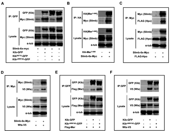
Figure 6—figure supplement 2. Complex formation and Kibra (Kib) degradation.

Figure 6—figure supplement 2.

(A–D) Slimb forms a complex with wild-type Kib and KibΔWW1&2 (A), Mer1-600 (B), Hpo (C), and Wts (D). (E–F) Co-IP of wild-type Kib or KibΔWW1&2 with Mer (E) or Wts (F). All experiments were performed using lysates from transfected S2 cells.