
FIGURE 1.
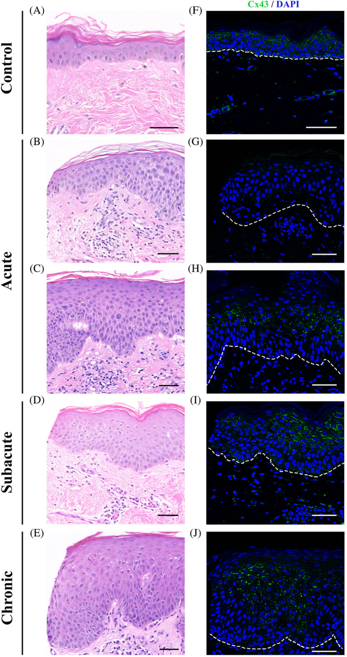
Downregulation of Cx43 expression in the spongiotic epidermis of eczema patients. Representative images of Cx43 staining of skin biopsy samples from normal skin (F) as well as patients with acute eczema (G, H; subject E10), subacute eczema (I; subject E6), and chronic eczema (J, subject E9). Corresponding H&E images of Cx43 staining (A‐E). Scale bars = 50 μm, 40× images. Dotted line marks the outline between the epidermis and dermis. Cx43 staining was visibly reduced in spongiotic areas, which are more commonly found in acute eczema samples. In subacute and chronic eczema samples Cx43 staining was closer to normal Cx43 expression shown in control skin except in regions where spongiosis could be observed