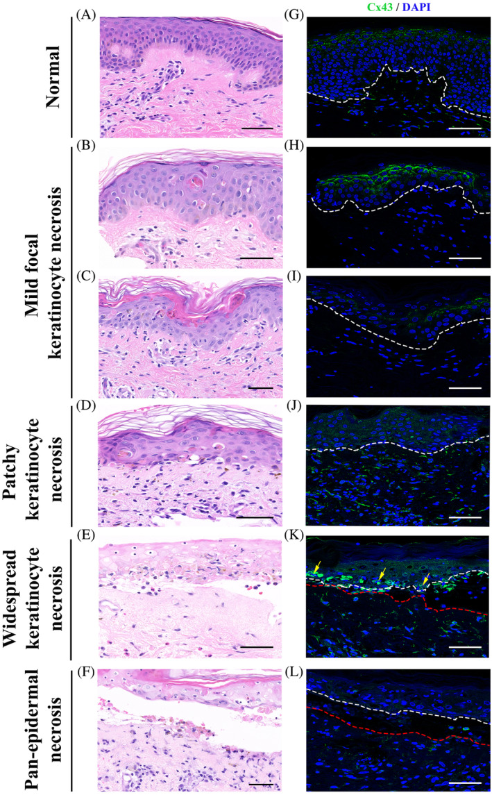
FIGURE 3.

Change in Cx43 expression in patients with SJS/TEN. Representative images of Cx43 staining of skin biopsy samples from normal skin (G) and patients with SJS or SJS/TEN overlap showing mild focal keratinocyte necrosis (H, I; from subject S8), patchy areas of keratinocyte necrosis (J; from subject S1), widespread keratinocyte necrosis (K; from subject S10), and pan‐epidermal necrosis (L; from subject S5). Corresponding H&E images of Cx43 staining (A‐F). Arrows mark infiltration of leucocytes. White and red dotted line marks the epidermis and dermis, respectively. Scale bars = 50 μm, 40× images. Cx43 expression is elevated in the epidermis showing focal keratinocyte necrosis. As the keratinocyte necrosis becomes more widespread in the epidermis, Cx43 expression becomes cytoplasmic compared to normal punctate staining in control skin. In the sections showing pan‐epidermal necrosis Cx43 expression is lost at areas of epidermal detachment
