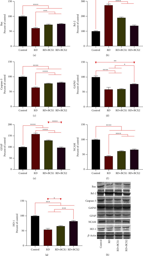
Figure 4.

Effects of BCX on retinal protein levels of Bax (a), Bcl-2 (b), Caspase-3 (c), Gap43 (d), GFAP (e), NCAM (f) and HO-1 (g) levels in rats with RD. The intensity of the western blot bands (h) was quantified by densitometric analysis, and β-actin was included to ensure equal protein loading. Data are expressed as a ratio of the control value (set to 100%). The bar represents the standard error of the mean. Blots were repeated at least three times (n = 3) and a representative blot is shown. Asterisks indicate statistical differences among groups (∗p < .05; ∗∗p < .01; ∗∗∗p < .001; ∗∗∗∗p < .0001). BCX: β-cryptoxanthin; Bax: Bcl-2-associated X; Bcl-2: B-cell lymphoma 2; Caspase-3: cysteine-aspartic acid protease-3; GAP43: growth-associated protein 43; GFAP: glial fibrillary acidic protein; HO-1: heme oxygenase-1; NCAM: neural cell adhesion molecule; NF-κB: nuclear factor kappa B; RD: retinal degeneration.
