
Figure 4.
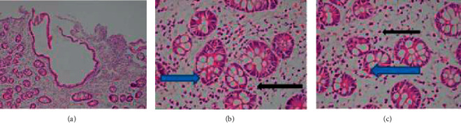
Histologic findings of duodenal biopsy; the duodenum mucosa shows (a) villous atrophy, architecture changes, and edematous lamina propria, (b) plasma cells (black arrow), and (c) cryptitis (blue arrow) in the lamina propria.
Official websites use .gov
A
.gov website belongs to an official
government organization in the United States.
Secure .gov websites use HTTPS
A lock (
) or https:// means you've safely
connected to the .gov website. Share sensitive
information only on official, secure websites.
Histologic findings of duodenal biopsy; the duodenum mucosa shows (a) villous atrophy, architecture changes, and edematous lamina propria, (b) plasma cells (black arrow), and (c) cryptitis (blue arrow) in the lamina propria.