
Fig. 2.
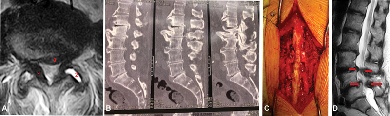
(A) Axial magnetic resonance imaging (MRI) showing spinal stenosis with ligamentum flavum thickening (1), facet arthrosis with synovitis (2) and disc protrusion (3). (B) Sagittal computed tomography (CT) scan showing L1-L2-L3-L4 spinal stenosis. (C) Surgical photography: three-level decompression using the conventional laminectomy technique plus lateral recess resection and bilateral foraminotomy. (D) Sagittal MRI showing degenerative L3-L4 and L4-L5 stenosis with degenerative spondylolisthesis in L4-L5.