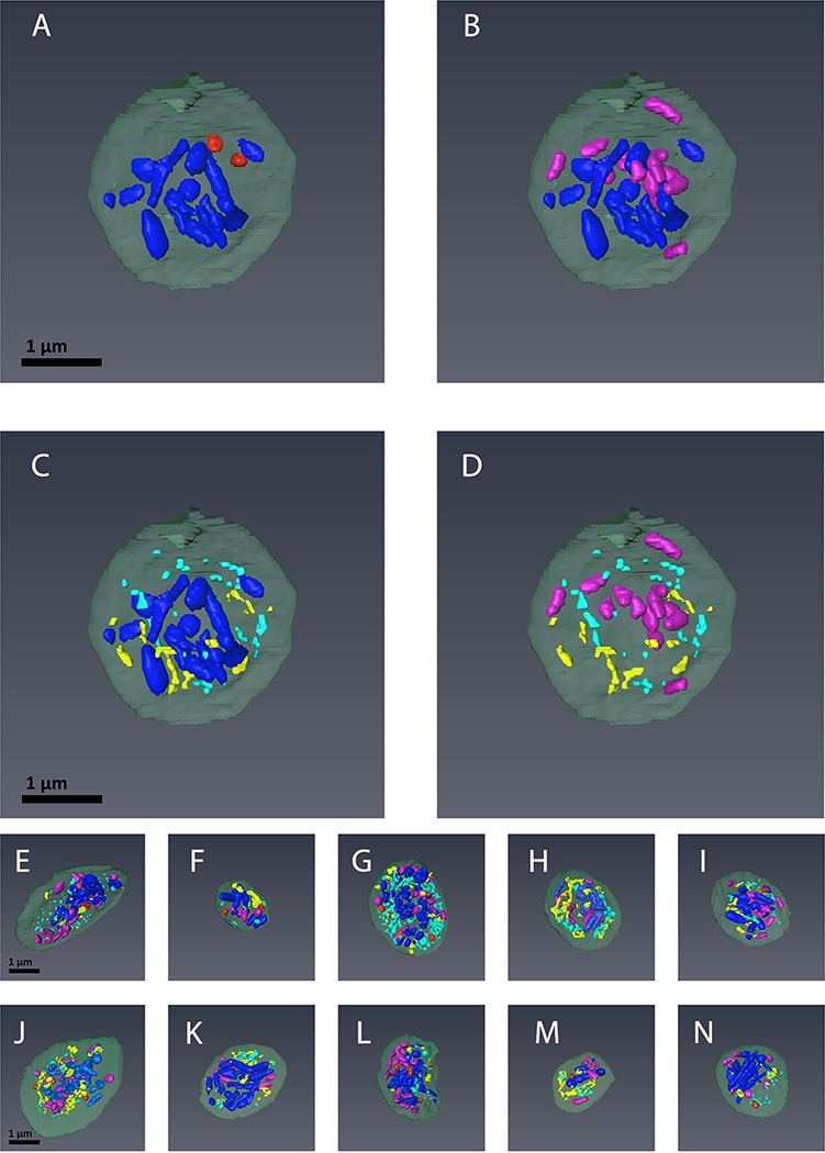
FIGURE 3. The spatial arrangement of α-granules, dense granules, canalicular system (open and closed), and mitochondria in mouse platelets.
Depicted are 3D renderings of individual platelets in which the organelles have been color-coded: α-granules (blue), dense granules (red), mitochondria (purple) and open and close canalicular system (yellow and turquoise, respectively). The relative distributions for given organelles are highlighted by direct comparison: A. α-Granules vs. Dense Granules; B. α-Granules vs. Mitochondria; C. α-Granules vs. Canalicular System; D. Mitochondria vs. Canalicular System. E-N. Depict combined color-coded images for 10 individual platelets rendered in 3D.

