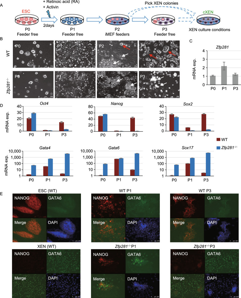
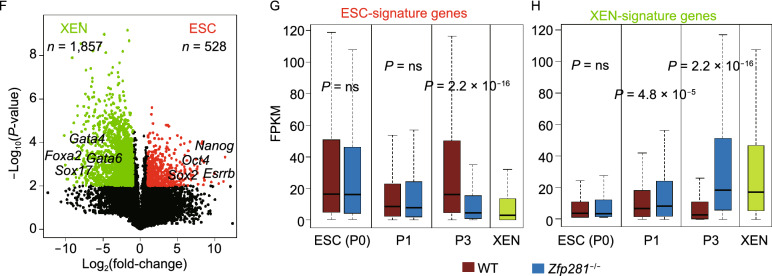
Figure 1.
ZFP281 functions as a barrier in ESC-to-XEN differentiation. (A) A schematic plot of ESC-to-XEN differentiation in vitro. To avoid contamination of irradiated MEF feeders, RNAs were collected at P0 (passages 0), P1 and P3 (feeder-free) for qRT-PCR analysis. (B) Phase contrast microscope images of WT and Zfp281−/− ESC-derived cXEN cells at P0-P3. White and red arrows indicate the XEN-like and ESC-like colonies, respectively. (C and D) qRT-PCR analysis of Zfp281 (C), pluripotency (Oct4, Nanog, Sox2) and PrE (Gata4, Gata6, Sox17) (D) transcripts in WT and Zfp281−/− ESC-derived cXENs at P0, P1, and P3. (E) Immunostaining of NANOG and GATA6 at P1 and P3 in ESC-to-XEN differentiation. WT ESCs and XENs were used as positive controls for NANOG and GATA6 staining, respectively. (F) A volcano plot of differentially expressed genes (DEGs, fold-change > 2, T-test P-value < 0.01) between ESCs and XENs. DEGs highly expressed in ESC and XEN were defined as ESC-signature genes and XEN-signature genes, respectively. (G and H) Box plots for the expression of ESC-signature genes (G) and XEN-signature genes (H) in WT and Zfp281−/− ESCs in cXEN differentiation. P-value was from a Mann-Whitney test


