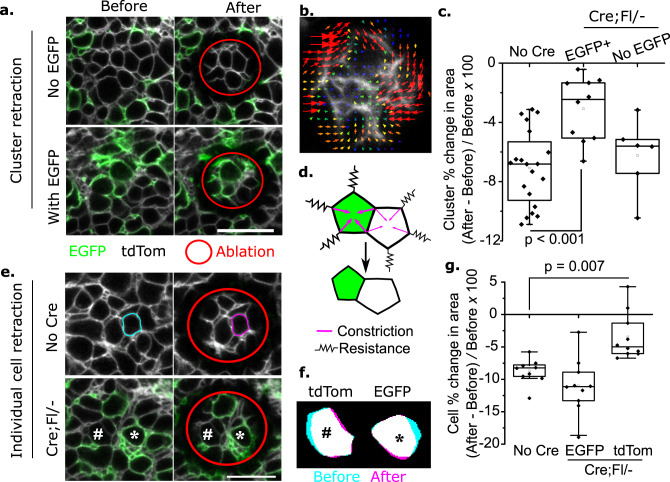
Fig. 3. Mosaic Vangl2 deletion non-autonomously diminishes neuroepithelial apical retraction.
a Representative (>6 embryos) live-imaged neuroepithelium before and after annular laser ablation. Ablations were made in regions including (with EGFP) or excluding (no EGFP) Vangl2-deleted cells. b Particle image velocimetry (arrows superimposed on grey cell borders) illustrating apical retraction of a cluster of cells within the ablated annulus. c Quantification of the retraction (% change in area) of cell clusters within the ablated annulus. Dots represent a cluster in an individual embryo; each embryo was only ablated once. Control n = 20, EGFP+ n = 10, No EGFP n = 6 embryos. d Schematic illustration of cell retraction due to immediate constriction when released from connections to surrounding cells. e, f Representative live-imaged neuroepithelium from control (No Cre, 20 embryos) and Cre;Fl/− (10 embryos). A cell is traced before and after ablation in the No Cre control. The # and * correspond to the cells shown in f, illustrating retraction. g Quantification of the retraction (% change in area) of individual cells within the ablated annulus. Ablations included 2–3 Vangl2-deleted (EGFP) and Vangl2-replete (tdTom) neighbours of the deleted cells. Retractions of cells of the same type were averaged in each cluster, n = 10 embryos per genotype. Scale bars = 20 µm. p values from ANOVA with post-hoc Bonferroni.

