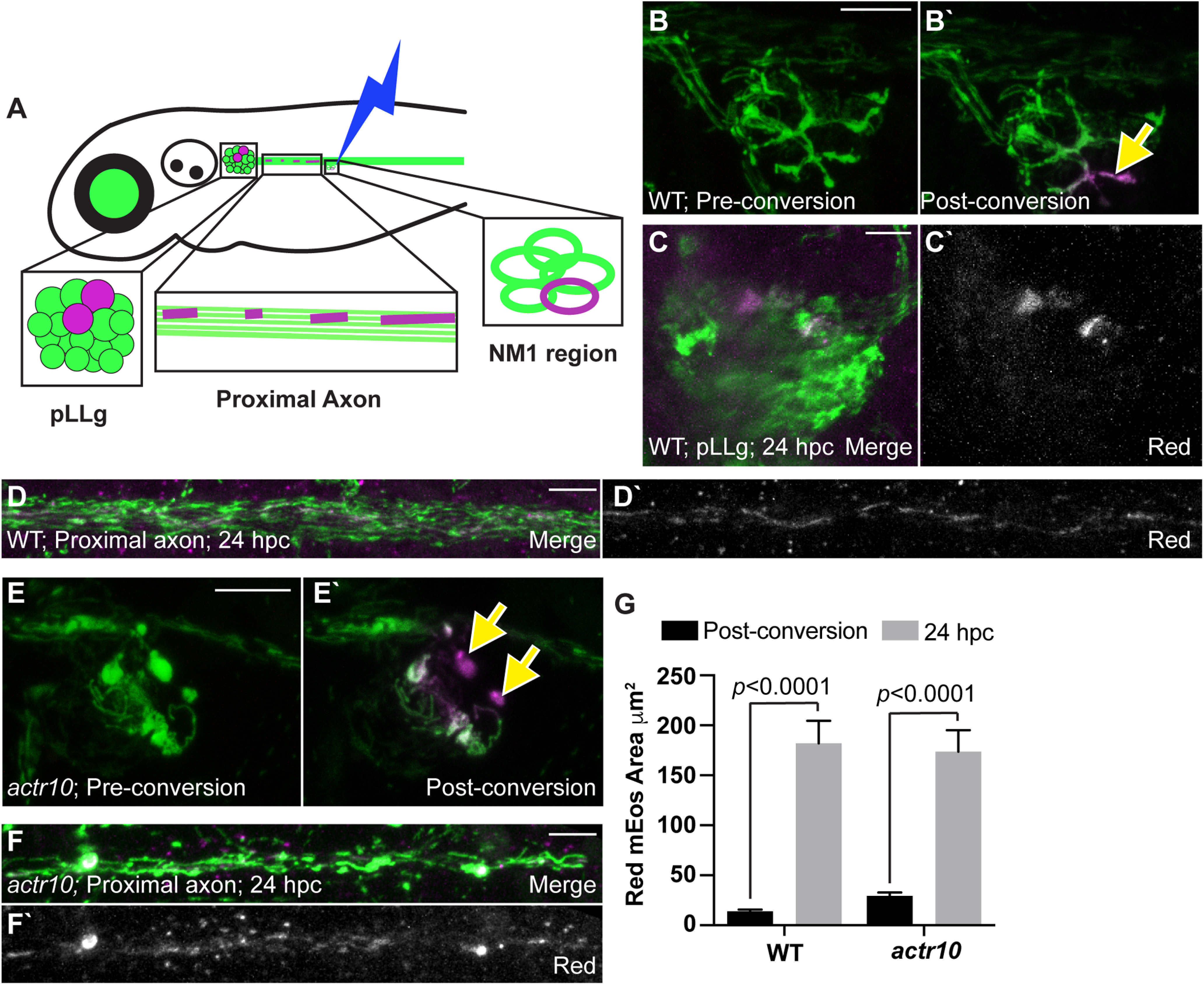
Figure 8.

mEos can be redistributed independent of retrograde transport in pLL neurons. A, Schematic of the minimal mitochondrial photoconversion strategy and the regions analyzed for mitochondrial area at 24 hpc. B, Images of a WT axon terminal before and immediately after photoconversion. Green: naive mEos; magenta: converted mEos. Arrow points to region photoconverted. C, D, The pLLg and proximal axon at 24 hpc shows mitochondrially localized converted (magenta in merge, white alone) mEos in the cell body and mitochondria along the axon. E, An actr10nl15 mutant axon terminal before and immediately after photoconversion. F, The proximal axon of an actr10nl15 mutant 24 hpc showing converted mEos in mitochondria in the axon. G, Quantification of the total area of converted mEos fluorescence immediately postconversion and at 24 hpc (ANOVA; WT: n = 9; actr10nl15: n = 13). Postconversion: WT: 14.16 ± 2.97; actr10nl15: 29.52 ± 2.60. 24 hpc: WT: 181.98 ± 23.70; actr10nl15: 173.55 ± 20.79. Scale bars: 10 µm. All data are mean ± SEM.
