
Figure 3.
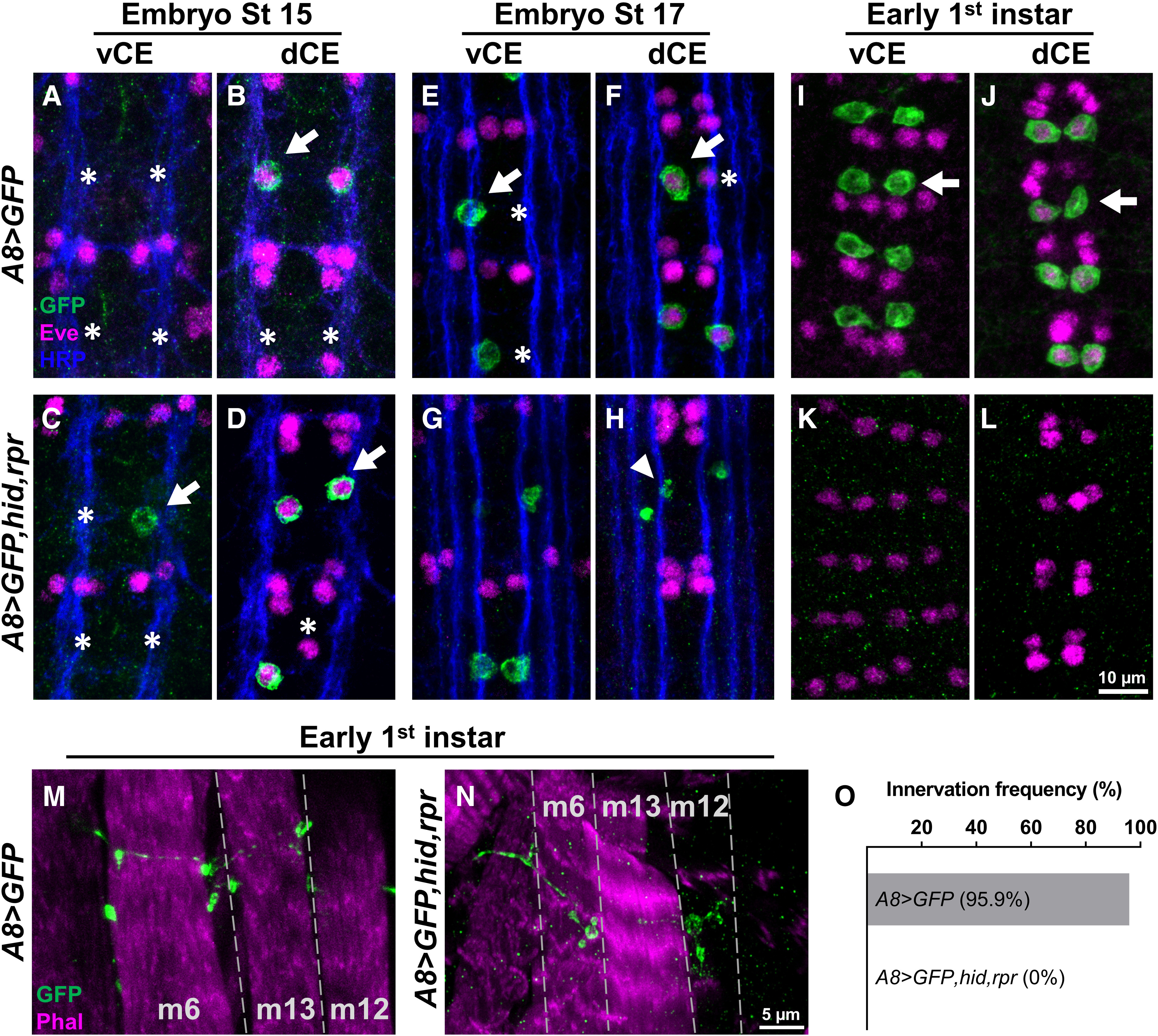
A8>hid,rpr-induced cell death occurs after 1s innervation. A–L, Representative images depicting the presence or absence of vCE and dCE cell bodies from embryonic stage 15 (A–D), stage 17 (E–H), and early first instar (I–L) larval VNCs labeled with GFP (green), Eve (magenta), and Fasciclin 2 (blue) in control (A8>GFP) and 1s-ablated (A8>GFP,hid,rpr) animals. Arrows and asterisks indicate the cells expressing or not expressing A8, respectively. A, B, A8 expression begins at embryonic stage 15. In A8>GFP,hid,rpr, vCEs, and dCEs undergo apoptosis starting at embryonic stage 17 (G, H), noted by the loss of Eve staining in dCE and GFP-positive debris (indicated by arrowhead), and are completely absent in early first instar larvae (K, L). M, N, Representative 1s NMJs labeled with GFP (green) and a muscle marker, phalloidin (magenta), in control (M) and 1s-ablated early first instar larvae (N). Note that 1s NMJs are labeled by GFP in control animals, and some 1s NMJs are still present in A8>GFP,hid,rpr animals, suggesting that A8>GFP,hid,rpr-induced cell death happens after 1s NMJ formation. O, Innervation frequency of 1s MNs in control and 1s-ablated late first instar larvae. Three muscles (m6, m12, and m4) were pooled and analyzed together. All 1s NMJs were eliminated in 1s-ablated animals by this stage. n values (NMJs/larva) are 76/5 and 86/5 for O.