Figure 2.
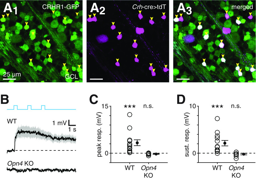
Rod- and cone-independent light responses of CRH+ ACs depend on melanopsin. A, Confocal micrographs showing GFP fluorescence (A1), tdT fluorescence driven by a Crh-cre allele (A2), and their overlap (A3) in the GCL of a Crhr1-gfp+;Crh-cre+/−;Ai14+/− retina. Arrowheads indicate small somas that exhibit overlap between both fluorescence channels. B, Effect of melanopsin KO on rod- and cone-independent light responses in CRH+ ACs. Top, Black trace represents mean rod- and cone-independent voltage response of presumed CRH+ ACs (n = 13) to photostimulation. Gray shading represents ± SEM across cells. Bottom, Same format as top but for presumed CRH+ ACs (n = 9) recorded in melanopsin KO (Opn4 KO; i.e., Crhr1-gfp+;Opn4Cre/Cre) retinas (Φstim = 1.5 × 10¹⁷ Q cm−2 s−1). C, D, Peak (C) and sustained (D) voltage responses in CRH+ ACs shown in B. ***p < 0.001.

