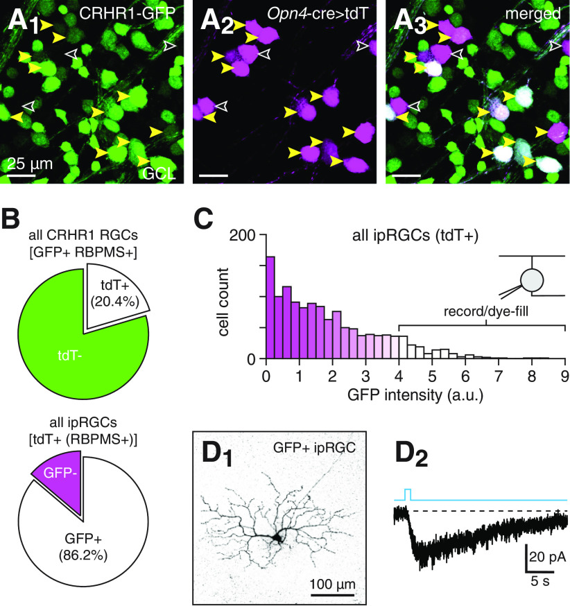
Figure 4.
Labeling of ipRGCs in Crhr1-gfp retinas. A, GFP labeling of a subset of genetically identified ipRGCs in a Crhr1-gfp+;Opn4Cre/+;Ai14+/− retina. Confocal micrographs show GFP fluorescence (green, A1), tdT expression driven by an Opn4-cre allele (Opn4-cre>tdT) (magenta, A2), and their overlap (A3) in the GCL of a Crhr1-gfp+;Opn4Cre/+;Ai14+/− retina. Yellow arrowheads indicate somas that exhibit overlap between both fluorescence channels. Empty arrowheads indicate tdT+ somas that lack GFP expression. B, Quantification of overlap between genetically labeled ipRGCs (tdT+) and GFP+ RGCs in the GCL of Crhr1-gfp+;Opn4Cre/+;Ai14+/− retinas; 20.4% of GFP+ RGCs are ipRGCs (top) and 86.2% of ipRGCs are GFP+ (bottom). C, GFP fluorescence intensity distribution of genetically labeled ipRGCs (tdT+) in Crhr1-gfp+;Opn4Cre/+;Ai14+/− retinas. D, Targeted recording and dye-filling of intensely GFP+ ipRGCs in Crhr1-gfp retinas. D1, Confocal micrograph of an intensely GFP+ ipRGC (>90th percentile in distribution shown in C) dye-filled during whole-cell recording in a whole-mount Crhr1-gfp retina. D2, Melanopsin-mediated intrinsic photocurrent of cell shown in D1 in response to a 1 s light pulse (Φstim = 1.5 × 10¹⁶ Q cm−2 s−1).

