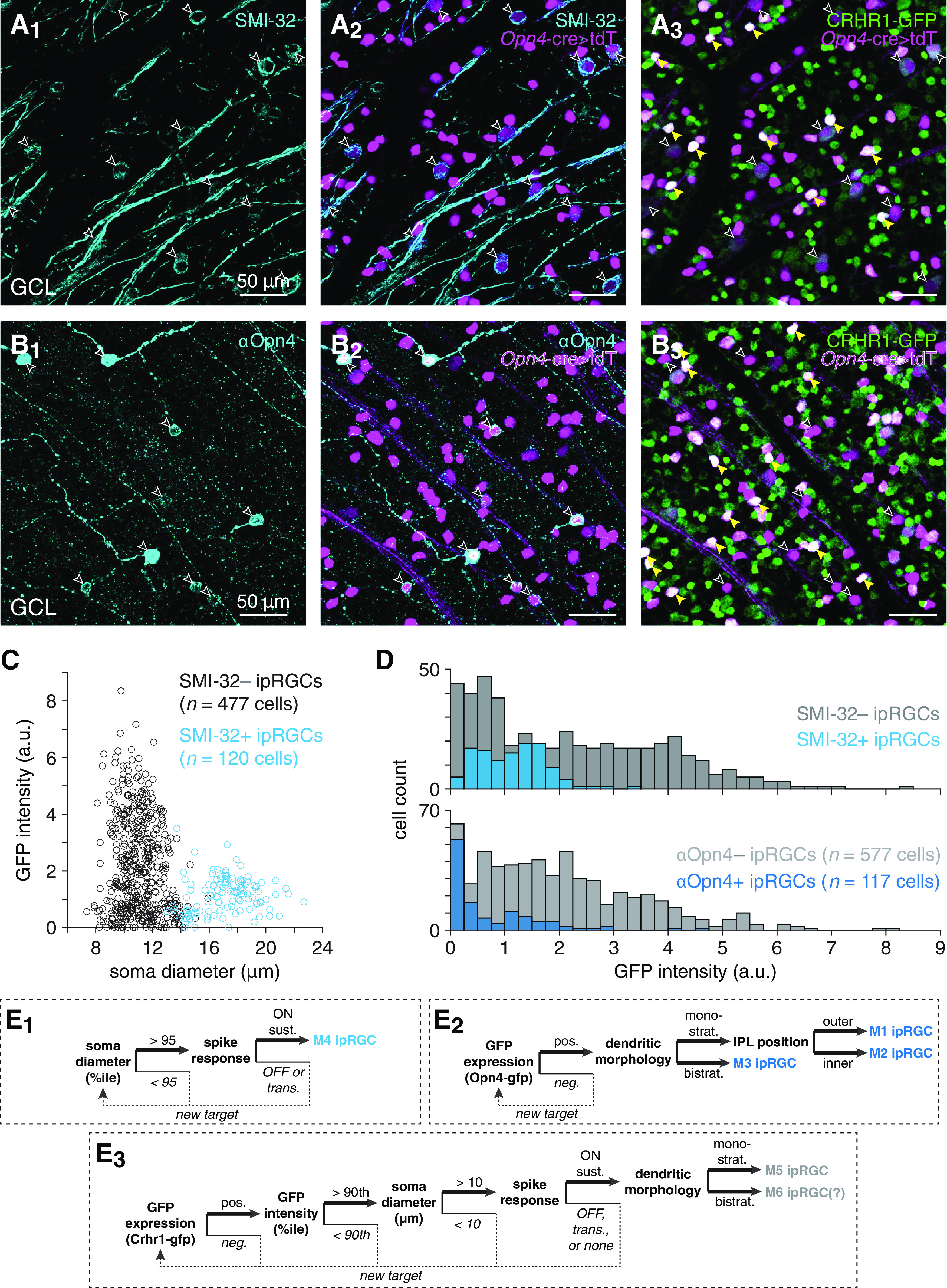
Figure 5.

Identification and targeting of ipRGC types labeled in Crhr1-gfp retinas. A, Visualizing overlap between SMI-32- and GFP-positive cell populations in Crhr1-gfp retinas. A1, Confocal micrograph showing SMI-32 immunostaining (cyan, A1) in the GCL of a Crhr1-gfp+;Opn4Cre/+;Ai14+/− retina. A2, same region as in A1, but also showing tdT expression driven by an Opn4-cre allele (Opn4-cre>tdT) (magenta). Empty arrowheads indicate somas dual-labeled by SMI-32 and tdT. A3, same as in A2, but showing GFP expression (green) instead of SMI-32 staining. Yellow arrowheads indicate strong overlap (white) between tdT and GFP fluorescence channels. B, Same format as in A, but for Crhr1-gfp+;Opn4Cre/+;Ai14+/− retina immunostained with an antibody against melanopsin (αOpn4). C, Soma size plotted against GFP fluorescence intensity for SMI-32+ and SMI-32– ipRGCs in Crhr1-gfp+;Opn4Cre/+;Ai14+/− retinas. D, GFP fluorescence intensity distributions of ipRGCs in Crhr1-gfp+;Opn4Cre/+;Ai14+/− retinas immunostained with SMI-32 (top) or αOpn4 (bottom). E, Decision trees for targeting specific ipRGC types: M4 (E1), M1-M3 (E2), and M5-M6 (E3).
