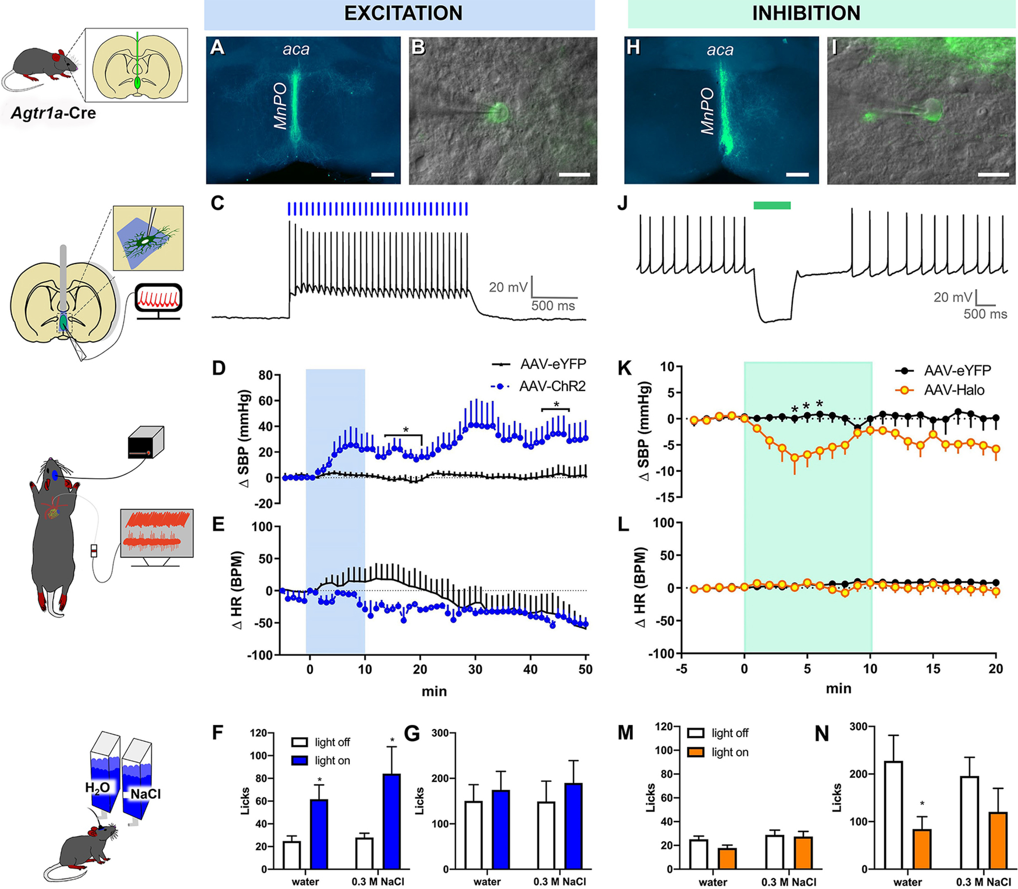
Figure 4.

Optogenetic stimulation of AT1aR-containing neurons in the MnPO/OVLT influences blood pressure. A, Cre-dependent expression of ChR2/eYFP (green) in MnPO/OVLT of an Agtr1a-Cre mouse. B, A combination of epifluorescence and DIC microscopy was used to target virally transformed neurons for study. C, Exposure to 15 Hz blue light produces action potentials. D, SBP and (E) heart rate (HR) response to blue light stimulation of AT1aR neurons residing in the MnPO/OVLT (10 mW; 15 Hz; 20 ms pulse width; 60 s ON/OFF; performed over a period of 10 min, n = 4 or 5/group). Water and 0.3 m NaCl intake in response to the same stimulation parameters (F) during free access to water and (G) subsequent to 16 h of water deprivation; n = 15 mice. H, Cre-dependent expression of halorhodopsin (Halo)/eYFP (green) in the MnPO/OVLT of an AT1aR-Cre mouse. I, A combination of epifluorescence and DIC microscopy was used to target virally transformed neurons for study. J, Green light hyperpolarizes AT1aR neurons in the MnPO/OVLT. Changes in (K) SBP and (L) HR in response to optogenetic inhibition of AT1aR neurons of the MnPO/OVLT in mice receiving AAV-Halo; n = 6/group. Water and 0.3 m NaCl intake in response to optogenetic inhibition (M) during free access to water and (N) subsequent to 16 h of water deprivation; n = 9 mice. aca, anterior commissure. Error bars indicate SEM. *p < 0.05. Scale bars: A, H, 250 µm; B, I, 20 µm.
