Figure 4.
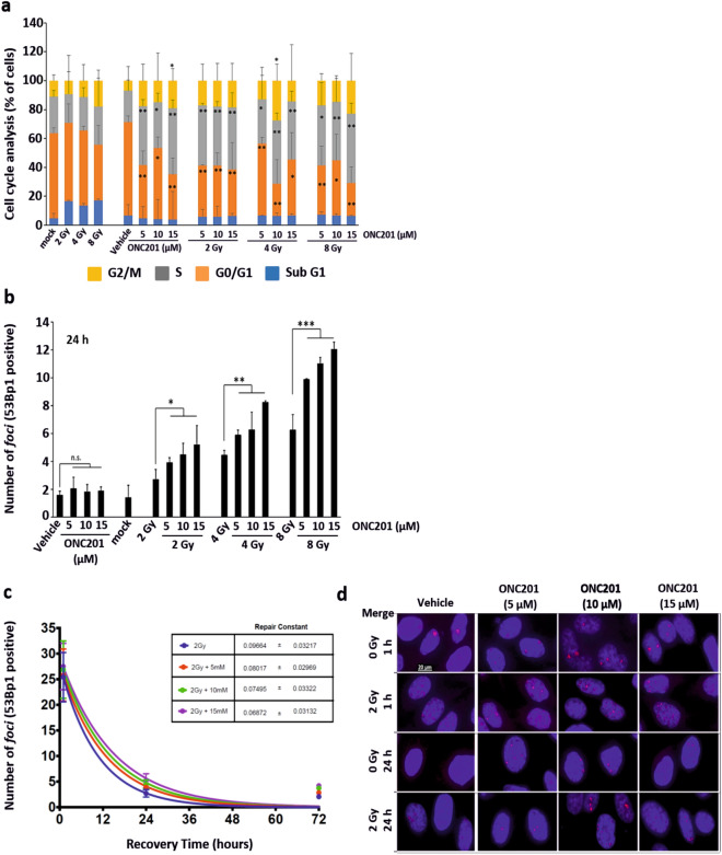
ONC201 determines the accumulation of foci into the nuclei of cells primed to radiation. (a) Cell cycle analysis of PC3 cells primed to radiation (Xrad) with ONC201 (5–15 µM) for 24 h showed an expansion of the cell population in S (grey) and G2/M (yellow) phases (n = 3). (b) Number of 53Bp1+ foci per cell at 24 h post-irradiation in PC3 cells. Samples were analysed at the time points schematically represented in Fig. 3a (n = 3). (c) Analysis of the kinetics of repair from the DNA damage induced by radiation in PC3 cells (n = 3). Samples were treated and analysed at the time points schematically represented in Fig. 3a. (d) Immunofluorescence analysis (Merge) of the foci formation, determined through 53Bp1 (red) staining at 1 and 24 h from radiation (representative of n = 3). The total number of foci counted is represented in panel (b). One-way ANOVA test has been run. The data shown in panel (b) have been run through ANOVA test on Ranks and further analysed with Dunnett’s Method. *p ≤ 0.05; **p < 0.01; ***p < 0.001 (± SD).

