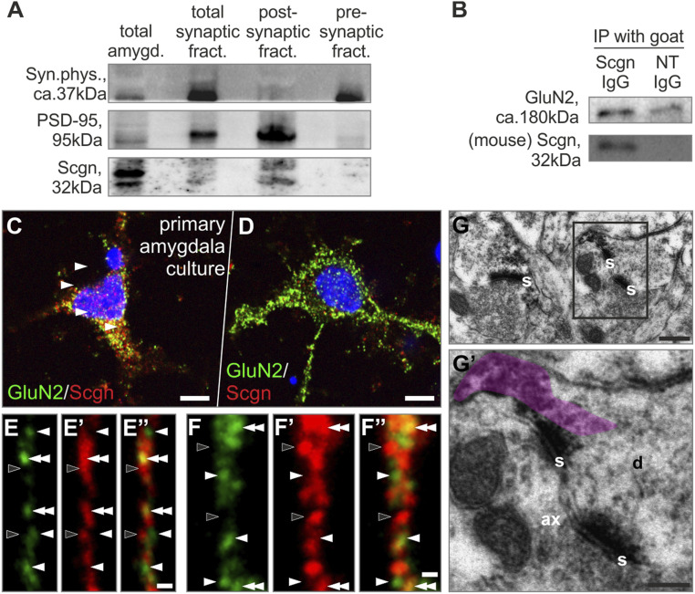
Fig. 3.
Secretagogin (Scgn) concentrates in the postsynaptic compartment of excitatory synapses. (A) Western blotting showed Scgn expression in the postsynaptic density fraction of amygdala punches. (B) GluN could be immunoprecipitated with an anti-Scgn antibody from amygdala punches. (C and D) GluN2+ neurons of primary amygdala culture could coexpress Scgn. (E–F’’) High-resolution airyscan imaging of GluN2+/Scgn+ neurons revealed close apposition of GluN2+ (white arrowheads) and Scgn+ (black arrowheads) puncta with colocalizations (double arrowheads). (G and G’) Scgn typically condensed in the postsynaptic compartment (immunoprecipitate semitransparently color-coded in violet). ax, axon; d, dendrite; GluN2, NMDA receptor 2 subunit; IP, immunoprecipitation; NT, IgG non-target immunoglobulin G; PSD-95, postsynaptic density protein 95; s, synapse. (Scale bars: C and D, 5 µm; E” and F”, 1 µm; G, 100 nm; G’, 70 nm.)

