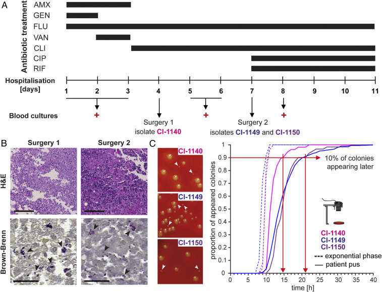
Fig. 1.
Clinical course, treatment, and lag-time analysis of S. aureus derived directly from a difficult-to-treat infection. (A) Overview of the clinical course, antibiotic treatment regimen, and surgical interventions. Positive blood cultures and isolated S. aureus strains are indicated. (B) Histological staining of patient tissue samples. Hematoxylin and eosin (H&E) staining (top panel) shows immune cell infiltration; Brown-Brenn staining (bottom panel) visualizes bacteria. (Scale bar for H&E, 100 µm; for Brown-Brenn, 20 µm.) (C) Representative images of colony radius heterogeneity of bacteria plated immediately after surgery (left panel). Proportion of appeared colonies over time, when plating bacteria directly from patient pus (continuous line) and upon recultivation in nutrient-rich medium to exponential phase (segmented line) (right panel). Red arrows indicate time point at which 90% of the colonies appeared.

