Fig. 2.
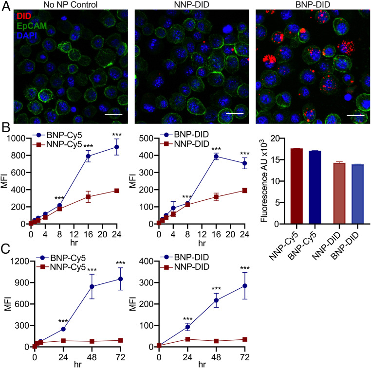
BNPs readily associate with tumor cells. (A) Confocal microscopy of PDV SCC cells following 24-h incubation with DiD dye-loaded NNPs and BNPs demonstrate superior affinity of BNPs to tumor cells. (Scale bar: 20 μm.) (B) Assessment of cellular association over time via flow cytometric analysis of PDV cultured under serum-free conditions with Cy5-conjugated NPs (Left) and DiD-loaded NPs (Center) demonstrates significantly improved association of BNP in both cases, with their advantage especially notable at later time points (16 to 24 h). Baseline fluorescence (Right) of both dye-conjugated (Cy5) and -loaded (DiD) NNP and BNP before incubation is similar. A comparison of NP association with tumor cells following short (up to 5 h) incubation in serum-containing vs. serum-free conditions is shown in SI Appendix, Fig. S2. (C) Cellular association of Cy5-NPs (Left) and DiD-NPs (Right) with extended incubation in serum-containing media. Even with prolonged exposure, BNPs associate with tumor cells more strongly than NNPs. MFI, mean fluorescence intensity; n = 3 replicates. ***P < 0.001, Student’s t test.

