Figure 5.
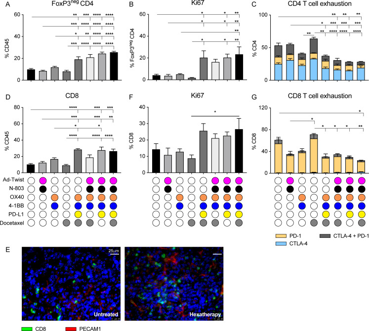
Treatment with the hexatherapy regimen results in tumor-infiltrating T cells that are more proliferative and less exhausted in the ‘cool’ 4T1 tumor model. Primary tumors (n=4–5/group) from figure 4B, C were collected on day 28 post-tumor implantation (7 days after the last treatment). (A–C) Flow cytometry was performed to determine (A) the frequency of FoxP3neg CD3+CD4+ T cells in the CD45+ tumor-infiltrating immune population, (B) the frequency of Ki67+ expression in the FoxP3neg CD4+ T cells, and the (C) frequency of CTLA-4 and PD-1 expression in the CD4+ T cells. (D) Likewise, flow cytometry was performed to determine the frequency of CD3+CD8+ T cells in the CD45+ compartment. (E) Immunofluorescence staining of CD8+ T cells (green) and PECAM1+ cells (red) was performed on untreated and hexatherapy regimen-treated tumors to further elucidate T cell infiltration. (F, G) Flow cytometry was done to assess (F) the frequency of Ki67+ expression, and the (G) frequency of CTLA-4 and PD-1 expression in the CD8+ T cells. Statistical test: Analysis of variance with Tukey’s post hoc test. Error bars, SEM. *p<0.05; **p<0.01; ***p<0.001; ****p<0.0001. PD-1, programmed cell death protein-1; PECAM1+, platelet endothelial cell adhesion molecule-1 positive.

