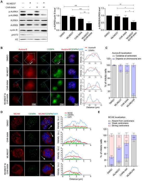
FIGURE 5.

Synergistic inhibition of Aurora‐A and Haspin disrupts the mitotic centromere aggregation of Aurora‐B and MCAK. A. The kinase activity of Aurora‐A and Aurora‐B was examined using Western blotting assays after releasing MDA‐MB‐231 cells from G2/M synchronization to indicated drugs for 30 min. The concentrations of MLN8237 and CHR‐6494 were 200 nmol/L. The repeated‐measures ANOVA, followed by the least significant difference test was used to evaluate the difference between groups. n = 3 independent experiments; ns, not significant; *P < 0.05; **P < 0.01. B. Left panel, immunofluorescence shows the location of Aurora‐B and CENPA. Right panel, the fluorescence intensity of Aurora‐B and CENPA was measured. C. The localization of Aurora‐B at centromere was analyzed (n = 3 independent experiments; ≥ 60 cells per condition). D. Immunofluorescence shows the location of MCAK. The arrows represent the direction of the line scan of fluorescence intensity for the entire cell. E. The localization of MCAK at the centromere was analyzed. “Strong” means that more than 50% of the centromeres show MCAK localization, while “weak” indicates that less than 50% of the centromeres show MCAK localization (n = 3 independent experiments; ≥ 123 cells per condition) Abbreviations: MLN, MLN8237; CHR, CHR‐6494; DAPI, 4', 6‐diamidino‐2‐phenylindole
