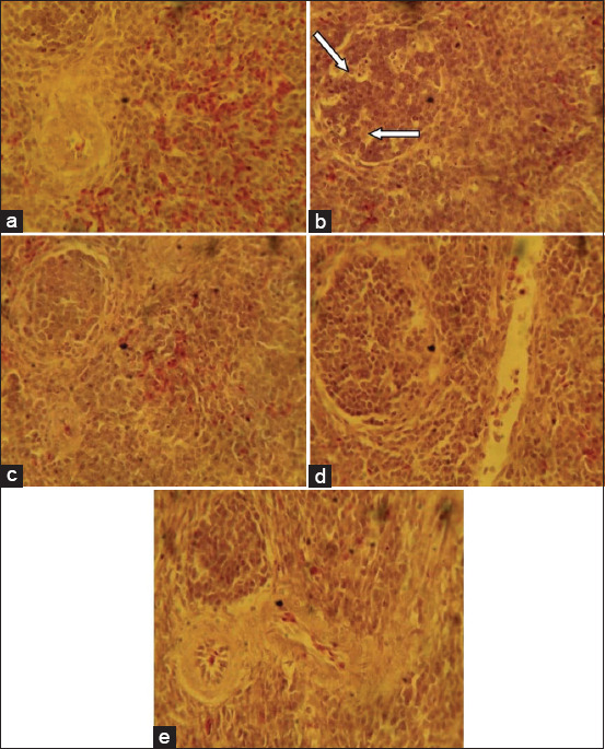
Figure-3.

Photomicrograph of spleen sections from the experimental groups: (a) Birds fed basal feed, showing normal histological features, (b) birds fed mold-contaminated feed, showing cellular depletion and degenerative changes in the white pulp of the germinal center (arrows), (c) birds fed mold-contaminated feed+Saccharomyces cerevisiae, (d) birds fed mold-contaminated feed+bentonite, and (e) birds fed Mold-contaminated feed+kaolin show no observable histological change (H and E, ×400).
