
Figure 1:
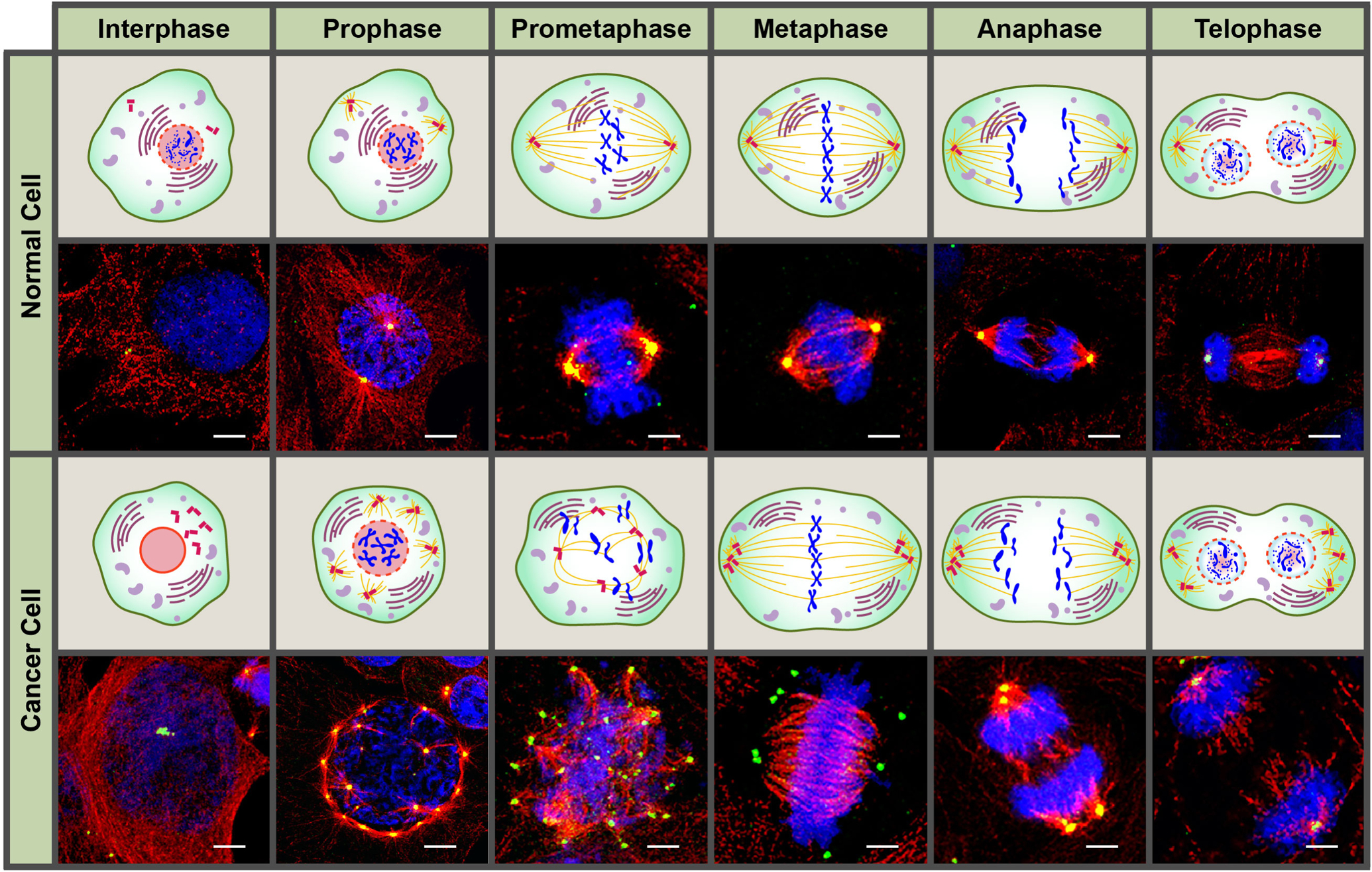
Confocal immunomicrographs and graphic illustration of a normal and cancer cell with extra centrosomes across the different phases of the cell cycle. Centrosomes and microtubules were immunolabeled for γ-tubulin (green) and α-tubulin (red), respectively, and DNA was counterstained with Hoechst (blue). Scale bar (white) = 5 μm.