
Fig. 1. Mortalin depletion effectively suppresses vemurafenib-resistant B-RafV600E melanoma cells.
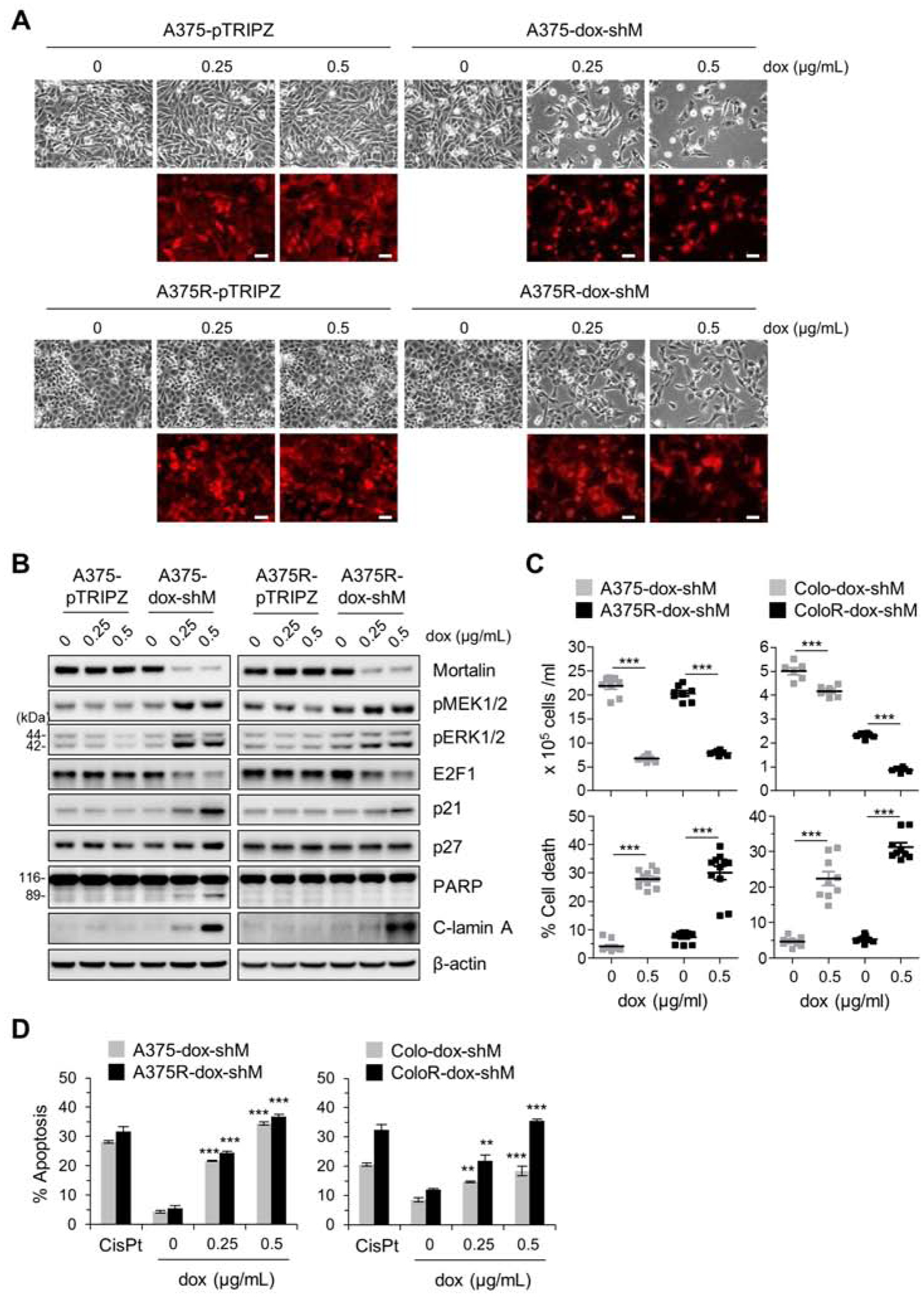
(A) Microscopic images of A375 and its vemurafenib-resistant progeny (A375R) stably expressing pTRIPZ-dox-shMort (A375-dox-shM and A375R-dox-shM, respectively) or the control pTRIPZ (A375-pTRIPZ and A375R-pTRIPZ, respectively). Cells were treated with doxycycline (dox) at indicated concentrations for 5 days. Red fluorescence indicates doxycycline-induced RFP expression and thus infection efficiency. Scale bar = 100 μm. (B) Western blot analyses of total lysates of cells treated as described in (A). C-lamin A indicates cleaved lamin A. β-actin is the control for equal protein loading. Densitometry of Western blots is shown in supplemental figure S1. (C) Cell viability determined by trypan blue exclusion assay after 5 day doxycycline treatment. Data are mean ± SEM from at least 6 independent experiments. (D) Apoptosis determined by Annexin V-FITC/TO-PRO 3 assay after 5 day doxycycline treatment. Colo-dox-shM and ColoR-dox-shM indicate Colo-829 and Colo829R stably expressing pTRIPZ-dox-shMort, respectively. Cells treated with 25 μM cisplatin (CisPt) for 24 hours are the positive control for apoptosis. Data are mean ± SEM, n = 3. Representative FACS histograms are shown in supplemental figure S2. P values are relative to untreated control. ** P < 0.01, and *** P < 0.001 by two-tailed paired student’s t test in (C) or by one-way ANOVA with Dunnett post-tests in (D).