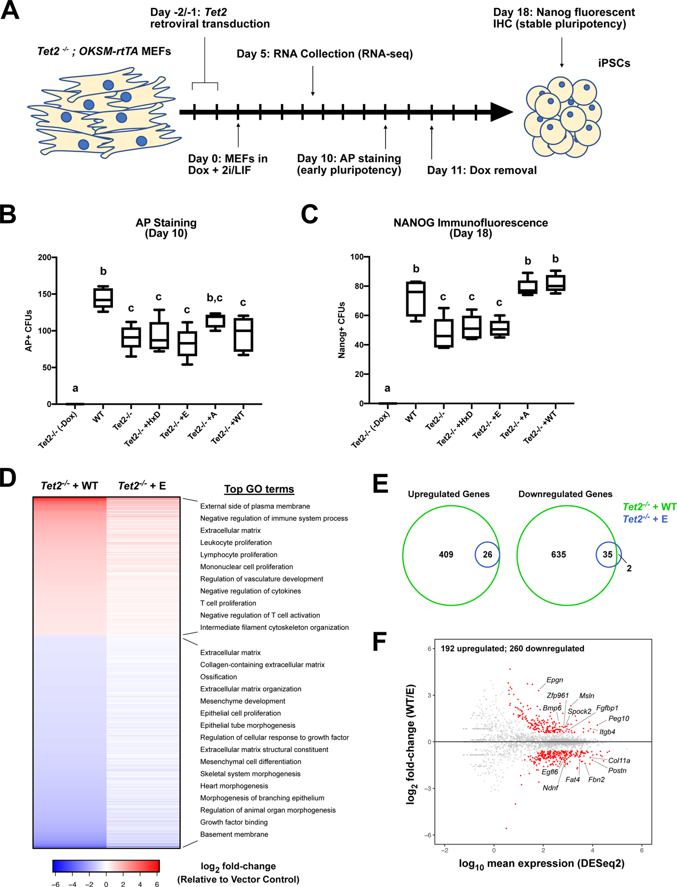
Figure 2 – Rescue of Tet2−/− iPSC reprogramming efficiency is dependent on TET2 fC/caC activity.

(A) Schematic illustration of Tet2−/− MEF reprogramming paradigm. (B) Quantification of AP staining of pluripotent colonies at day 10. Box plots indicate median AP+ colony forming units (CFUs) (one-way ANOVA with Tukey multiple comparisons; n=5; groups with different letters denote significant differences between groups, while shared letters indicate no difference was detected). (C) Quantification of immunohistochemical staining for NANOG-positive stable pluripotent colonies at day 18 (median NANOG+ CFU; one-way ANOVA with Tukey multiple comparisons; n=5). (D) Heat map of differentially expressed genes in Tet2-WT or Tet2-E transduced cells relative to vector control at day 5, with enriched GO terms indicated on right (RNA-seq; n=3–4; FDR < 0.05; fold-change > 1.5). (E) Venn overlap of significantly altered transcripts in Tet2-WT and Tet2-E transduced cells relative to vector control. (F) MA plot for Tet2-WT vs. Tet2-E gene expression. Red dots represent differentially expressed genes, with biologically relevant genes designated (FDR < 0.05; fold-change > 1.5).
