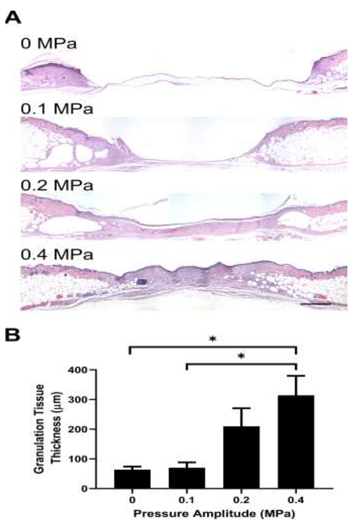
Fig. 2. Effects of pulsed ultrasound on granulation tissue deposition two weeks after injury.

Full thickness punch biopsy wounds were either sham-exposed or exposed to 1-MHz, pulsed ultrasound at pressure amplitudes of 0.1, 0.2, or 0.4 MPa for 2 weeks. Fourteen days after injury, animals were sacrificed, and wounds were excised and processed for histology. (A) H&E-stained skin sections from the center of wounds exposed at 0, 0.1, 0.2 or 0.4 MPa ultrasound. Images represent average granulation tissue thickness values from each group. Scale bar = 500 μm. (B) Granulation tissue thickness was measured at the wound center and plotted as an average for each treatment condition (n = 11-19). * p < 0.05 using the Kruskal–Wallis test with Dunn’s post-hoc test. Significant differences found between 0.4 MPa and 0 MPa (p = 0.026) as well as 0.4 MPa and 0.1 MPa (p = 0.017). Error bars indicate SEM.
