
Fig. 4. Quantification of granulation tissue deposition in diabetic wounds after 3 weeks of ultrasound treatment.
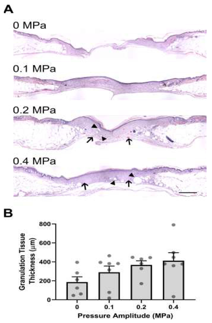
Full-thickness, diabetic mouse wounds were exposed to 1-MHz pulsed ultrasound at 0, 0.1, 0.2, or 0.4 MPa for 3 weeks. Three weeks after injury, mice were sacrificed, and their wound tissue harvested. (A) H&E-stained skin sections from the center of wounds. Images represent average granulation tissue thickness values from each group. Scale bar = 500 μm. (B) Granulation tissue thicknesses at the center of the wounds were quantified (n = 6-7). Graph shows individual data points representing the granulation tissue thickness of each animal, bars represent mean values for each treatment condition, and error bars indicate SEM.