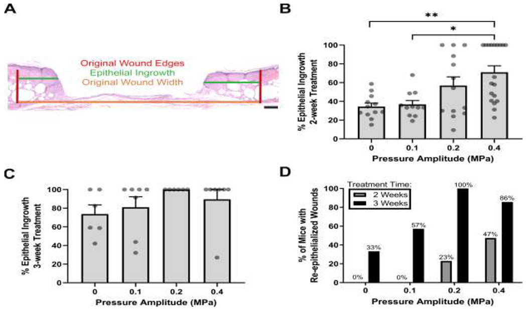
Fig. 6. Effects of ultrasound exposure on wound re-epithelialization.

Tissue sections corresponding to wound centers were used to measure re-epithelialization after 2 or 3 weeks of ultrasound exposure. (A) Representative image of H&E-stained skin section illustrating measurements used to quantify re-epithelization. Shown are the original wound edges (red lines), epithelial ingrowth (green lines), and original wound width (orange line). Scale bar = 200 μm. Epithelial ingrowth was measured and reported as a percent of the original wound width at (B) 2 weeks (n = 11-19) and (C) 3 weeks (n = 6-7) after injury. Circles represent data from individual animals and bars indicate mean values (± SEM). Significance tested (*p < 0.05 and **p < 0.01) by Kruskal–Wallis test and Dunn’s post-hoc test. Significant differences were found at 2 weeks between 0.4 MPa and both 0 MPa (p = 0.006) and 0.1 MPa (p = 0.022) conditions. (D) Measurements of epithelial ingrowth were used to calculate the percent of mice that had fully re-epithelialized wounds in each experimental group. Data are plotted as a function of pressure amplitude for 2-week (gray) and 3-week (black) exposures.
