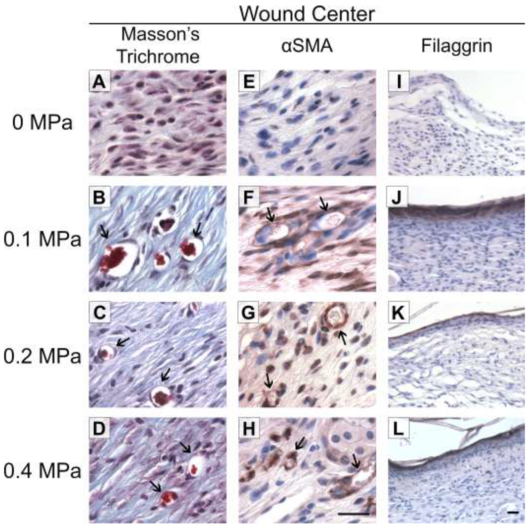
Fig. 7. Re-vascularization and epidermal maturation of ultrasound-exposed wounds.

Full-thickness wounds were either sham-exposed (A, E, I) or exposed to 1-MHz ultrasound at pressure amplitudes of 0.1 MPa (B, F, J), 0.2 MPa (C, G, K), or 0.4 MPa (D, H, L) for 3 weeks. Tissue sections from wound centers were stained with Masson’s trichrome (A-D) or anti-alpha-smooth muscle actin (αSMA) antibodies (E-H; brown staining) to visualize blood vessels (arrows). Scale bar = 25 αm. (I-L) Tissue sections from wound centers were stained with anti-filaggrin antibodies (brown). Scale bar = 25 αm.
