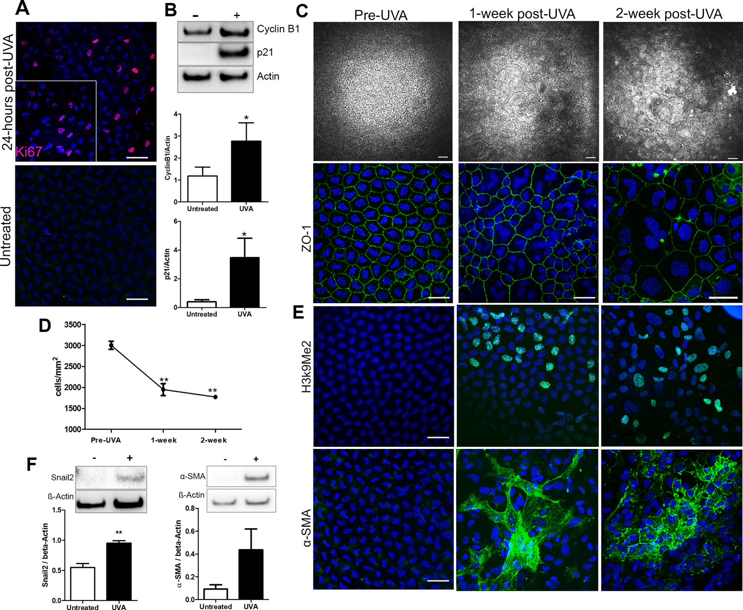
Figure 4.

UVA light activates the cell cycle leading to senescence and fibrosis in vivo. A. Ki67 expression in mouse CEnCs 24-hours post-UVA irradiation (top) and untreated (bottom). B. Representative western blots and densitometry analysis of Cyclin B1 and p21 levels in mouse CEnCs untreated vs 24-hours post UVA irradiation (n=5). *P < 0.05. C. Progressive changes in mouse CEnC morphology from untreated (Pre-UVA) to 1- and 2-week post-UVA irradiation. Top panel displays HRT images, bottom panel shows ZO-1 staining. D. Cell density analysis from pre-UVA to 2-weeks post-UVA (n=3). **P<0.001 comparison between post-UVA groups and pre-UVA. E. H3k9Me2 (top) and α-SMA (bottom) staining in mouse CEnCs in untreated eyes, 1-, and 2-weeks post-UVA irradiation. F. Representative western blots and densitometry analysis of Snail2 and α- SMA levels in mouse CEnCs untreated vs 2-weeks post-UVA irradiation (n=5). **P < 0.001. Scale bars = 50µm.
