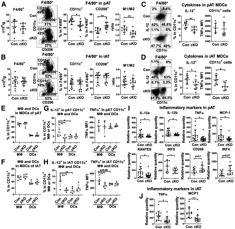
Figure 3.
cKO mice on HFD exhibit an improved macrophage phenotype in pAT and iAT. Male cKO mice and littermate controls were fed HFD for 16 weeks. Flow cytometric analysis of F4/80+ macrophages and DCs in SVCs isolated from pAT (A and C) and iAT (B and D) of cKO and control mice showing total F4/80+ cells, percentages of CD11c+ (M1-like MDC) and CD206+ (M2-like macrophage) cells in F4/80+ cells, ratio of CD11c+ to CD206+ cells (A and B), and intracellular levels of IL-12 and TNF-α in CD11c+ (F4/80+) MDCs (C and D). Flow cytometric analysis of CD11c+CD64+ macrophages and CD11c+CD64− DCs with CD11c+ MDCs of SVCs from pAT (E and G) and iAT (F and H) of cKO and control mice showing proportions of macrophages and DCs within MDCs (E and F) and intracellular levels of IL-12 and TNF-α in CD11c+ macrophages and DCs (G and H). RT-qPCR gene expression analysis of pAT (I) and iAT (J) of cKO and control mice showing mRNA levels of M1- and M2-like markers and cytokines. *P < 0.05, **P < 0.01. Con, control; MFI, mean fluorescence intensity.

