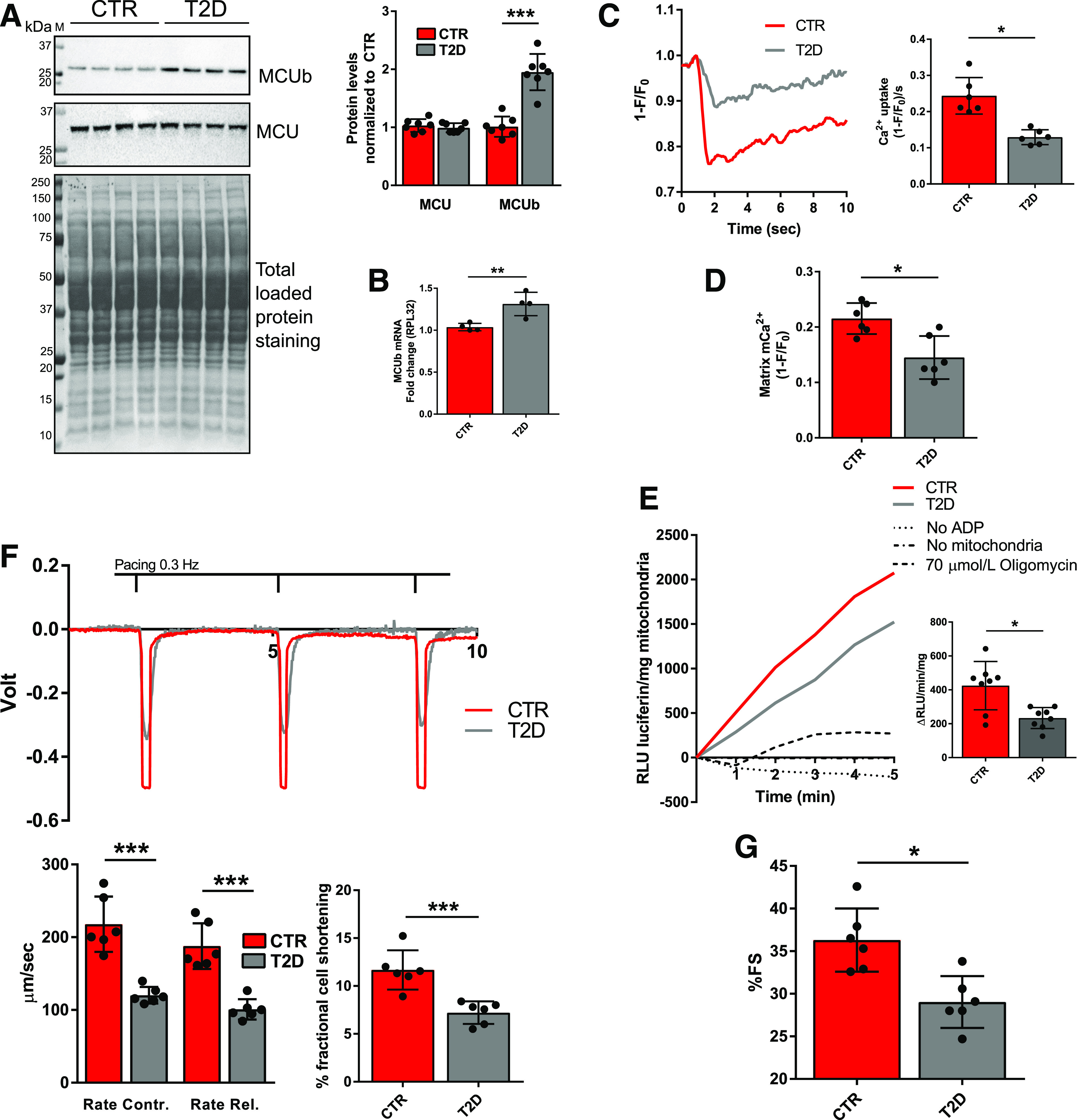
Figure 2.

MCUb overexpression in the T2D heart is associated with decreased mCa2+ import rate and level, decreased mitochondrial ATP production, and decreased cardiac function. A: Western blot analysis of MCUb and MCU in isolated cardiomyocyte homogenates. Total protein staining of polyvinylidene difluoride membranes was used as loading control. Summarized densitometric band analysis is shown. Western blots are representative of cardiomyocyte preparations obtained from seven mice per group. B: RT-qPCR analysis of MCU mRNA levels in CTR and T2D mouse hearts (four per group). 60S ribosomal protein L32 (RPL32) was used as the housekeeping gene for normalization. C: Representative mCa2+ transients measured in isolated cardiomyocytes from CTR and T2D. mCa2+ transients were assessed in paced-contracting cardiomyocytes (0.3 Hz) using MityCAM, which was delivered in vivo by injection of AAV-MityCAM. mCa2+ uptake rates were quantified and are reported in the bar graph as averaged maximal drop of MityCAM fluorescence (F) induced by electrical stimulation over normalized stable MityCAM fluorescence (F0 = 1) at time zero prior to initiation of electrical stimulation per second ([1 − F/F0]/s). Data are representative of recordings from cells isolated from six mice per group. D: mCa2+ levels reported as averaged maximal drop of MityCAM fluorescence (F) induced by electrical stimulation over stable MityCAM fluorescence (F0 = 1) at time zero prior to initiation of electrical stimulation (1 − F/F0). Data are representative of recordings from cells isolated from six mice per group. E: ATP production, expressed as the rate of linear increase of luciferin luminescence (RLU luciferin) per milligram of mitochondria. Rates were analyzed in mitochondrial preparations from eight mice per group in the presence of 5 mmol/L ADP, 0.15 mg/mL d-luciferin, and 1 μg/mL recombinant firefly luciferase. As negative controls, the assays were conducted in absence of either mitochondria or ADP or in the presence of 70 μmol/L of the ATP synthase inhibitor oligomycin. F: Cardiomyocyte contractility was assessed in vitro by edge detection and analyzed with Felix32 software. Rate of contraction (Contr.) (+dL/dt) and rate of relaxation (Rel.) (−dL/dt) are reported in micrometers per second upon conversion of volts (V) into millimeters (mm). Percentage fractional cell shortening was calculated as (ΔV[Vmax − V0]/cell size [mm]) ∗ 100. Data are representative of recordings from paced-contracting (0.3 Hz) isolated cells from six mice per group. G: In vivo heart function was assessed by M-mode echocardiography; percentage of fractional shortening (%FS) is shown. Data are representative of six mice per group. Data are presented as mean ± SD. Unpaired Student t test was used for statistical analysis of A and B. One-way ANOVA followed by Dunnett multiple-comparisons test was used for statistical analysis in C–G. *Adjusted P < 0.05; **P < 0.01; ***P < 0.001. M, molecular weight marker.
