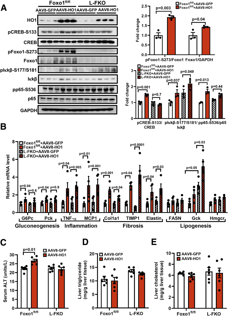
Figure 4.
HO1 overexpression in vivo activates NF-κB and Foxo1 in the liver. AAV8-GFP or AAV8-HO1 was delivered via retro-orbital to male mice (8–12 weeks old). Tissues from the mice were collected 14 days after the AAV8 injection. A: Total proteins from liver were extracted and loaded onto a 10% SDS-PAGE for Western blotting analyses to detect HO1, pCREB-S133, total CREB, pFoxo1-S273, total Foxo1, pIκkβ-S177/S181, total Iκkβ, pp65-S536, and total p65. The levels of phosphorylated proteins were normalized to the corresponding total protein level. The level of total Foxo1 was normalized to GAPDH. n = 3 mice/group. Results are presented as mean ± SEM. B: Total RNA from liver tissues were extracted for reverse transcription and quantitative PCR analyses of gluconeogenic, inflammatory, fibrogenic, and lipogenic genes. The relative mRNA level was normalized to cyclophilin. n = 4 mice/group. Results are presented as mean ± SEM. C–E: Blood samples from the mice were collected for the analysis of ALT activity, and liver tissues were collected for the analyses of triglyceride and cholesterol levels using commercial kits. n = 6 mice/group. Results are presented as mean ± SEM.

